In this interview, industry expert Jade Marrow discusses how NIR-II imaging supports deeper, higher-contrast preclinical imaging and helps researchers improve translational studies, biodistribution analysis, and real-time monitoring of biological processes.
To get started, could you give us a brief introduction to Scintica and explain how the company supports life sciences researchers globally, particularly in preclinical imaging and translational research?
Scintica is a global life sciences partner that works with multiple equipment manufacturers to distribute high-quality instrumentation to laboratories worldwide. Our products span a range of applications and research needs, including (but not limited to) optical imaging, ultrasound, MRI, SPECT, X-ray, and PET.
Among our diverse portfolio of modalities, we are committed to finding the best solution for your work. Our customers include those from academia, biotechnology companies, CROs, and government agencies. Scintica manages all aspects of the process, including marketing efforts, sales, procurement, equipment installation, system training, and post-sale support (either on-site or virtually). We provide technical and application expertise to help scientists every step of the way, going beyond the distributory role.
For researchers who may be familiar with traditional optical imaging but less familiar with NIR-II (SWIR) imaging, can you explain what NIR-II imaging is and where it fits within the broader imaging landscape?
Fluorescence imaging (FLI) in the second near-infrared (NIR-II) window, also known as short-wave infrared (SWIR), refers to wavelengths between 900 and 2000 nm, extending traditional FLI in the visible and NIR-I regions. The NIR-II range has many advantages for preclinical optical imaging because of its unique light-propagation physics, which enables greater penetration depth into biological tissues and lower autofluorescence due to reduced photon scattering and absorption.
Therefore, NIR-II supports higher-sensitivity imaging and offers clearer visualization of deep structures in an animal. Within the broader imaging landscape, NIR-II does not replace BLI or FLI in VIS and NIR-I but rather complements them by addressing their limitations in depth and spatial resolution.
The Newton FT-900 is designed to address this need, allowing researchers to perform sensitive BLI and conventional FLI alongside NIR-II imaging in a single all-in-one platform. This means users will not have to compromise their established workflows to gain access to this emerging optical technique.
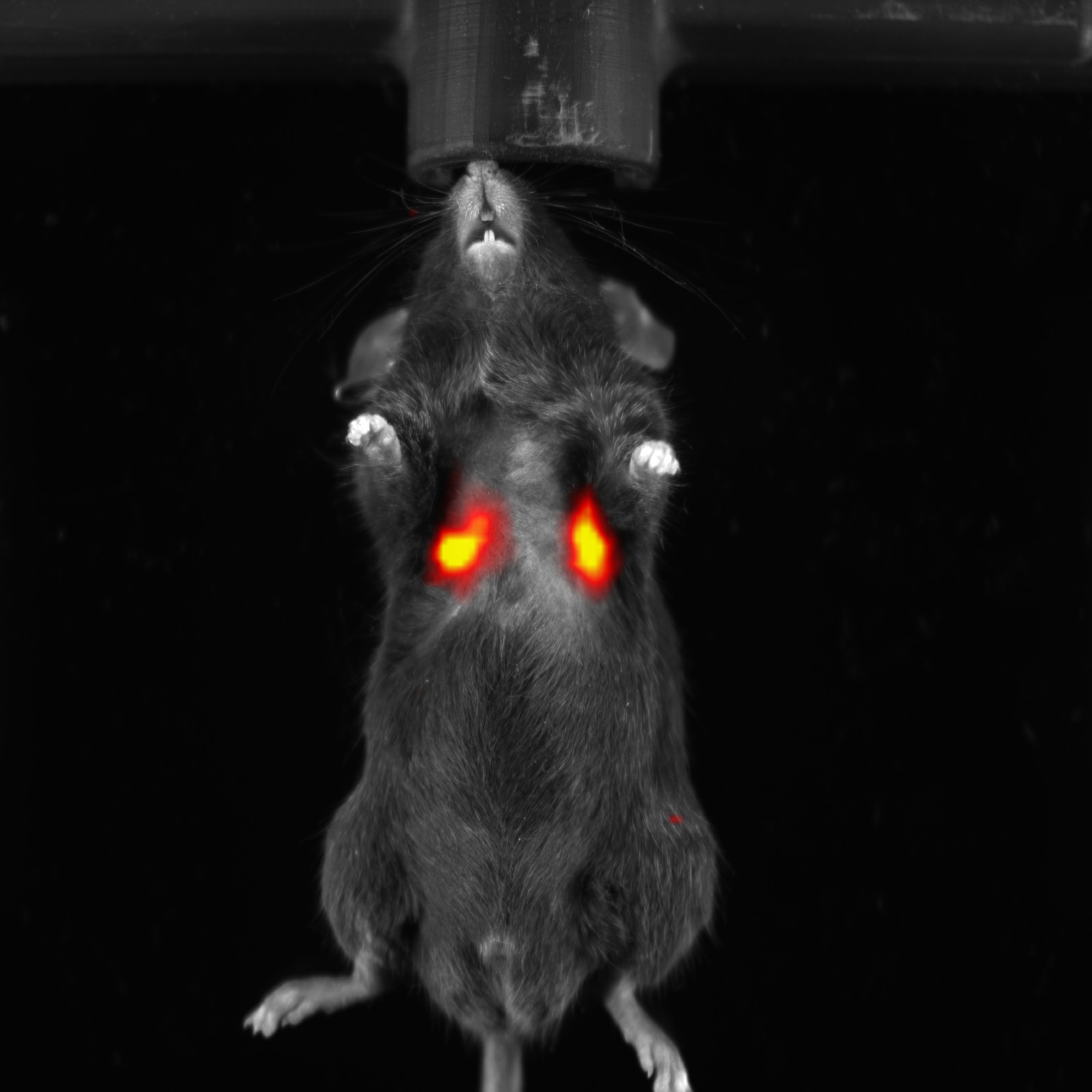
Image Credit: Scintica Instrumentation Inc.
What are the main limitations of conventional optical imaging methods, such as visible fluorescence or NIR-I fluorescence, that NIR-II imaging is designed to address?
Visible and NIR-I fluorescence are limited in terms of penetration depth, autofluorescence, photon scattering, sensitivity, and resolution. Wavelengths of light determine the degree of absorption by chromophores (e.g., hemoglobin, melanin, H2O, and lipids) and scattering in tissue (e.g., in brain, cranial bone, skin, and muscle); while these methods help to localize a signal well when it is just below the surface, they are less efficient when it is deep within the organism.
In comparison, NIR-II wavelengths travel more efficiently through biological tissues, experiencing less photon scattering and autofluorescence, which collectively enhance the signal-to-background ratio. In the NIR-II spectrum, the reduced absorption of emission signals by tissues helps improve spatial resolution and depth penetration, which can be up to 10x better than with visible fluorescence.
In practical terms, when should a researcher consider moving from BLI or NIR-I fluorescence imaging to NIR-II imaging?
A researcher may wish to transition from BLI or VIS/NIR-I FLI when they are investigating deep-tissue organs (e.g., liver, pancreas, brain, and vasculature), deep orthotopic tumors, and/or are using larger animal models (e.g., rats or obese mice). These deep applications are susceptible to greater light scattering, which limits the detectability of the true signal.
Further, those studying biodistribution and real-time pharmacokinetics benefit from using NIR-II, as its lower background noise and higher signal-to-noise ratio enable clearer visualization of drug accumulation and washout kinetics.
NIR-II may not be necessary in some cases, including when a study has already employed a BLI-based reporting system to evaluate promoter activity or gene expression; when the signal is superficial/subcutaneous; when the work involves a high-throughput assay focused solely on viability and no deep visualization is required; or when the experimental workflow is well-established and validated using VIS/NIR-I probes to achieve sufficient depth.
It is worthwhile noting that there is an entire field dedicated to designing and constructing next-generation probes for NIR-II. In the meantime, several commonly used and commercially available fluorophores, such as ICG and IRDye 800CW, can be used for NIR-II applications because they exhibit a second emission tail that extends into the 1000–2000 nm window.
Which preclinical applications are especially well-suited to NIR-II imaging, and why?
A) Deep-tissue imaging: VIS and NIR-I light scatter more across biological tissues (e.g., brain, bone, skin, and muscle), and the degree of VIS and NIR-I light absorption by chromophores (e.g., hemoglobin, melanin, H2O, and lipids) is greater compared to NIR-II.
B) Real-time drug distribution, pharmacokinetics, and metabolic imaging: NIR-II light allows for reduced background noise and the ability to track in real time to see drug accumulation and metabolism in deep tissues, such as the liver.
C) Cardiovascular mapping: Less light scattering and absorption by hemoglobin allow for the visualization of vascular architecture and perfusion. Cardiac and cerebral blood flow patterns and real-time vascular dynamics can be assessed using contrast agents.
D) Probe development and validation: There is an increasing need for commercially available NIR-II fluorophores. The Newton FT-900 enables proper evaluation of probe radiance, target specificity, and metabolism/clearance. This includes small molecule dyes, quantum dots, rare-earth nanoparticles, organic carbon dots, and single-walled carbon nanotubes.
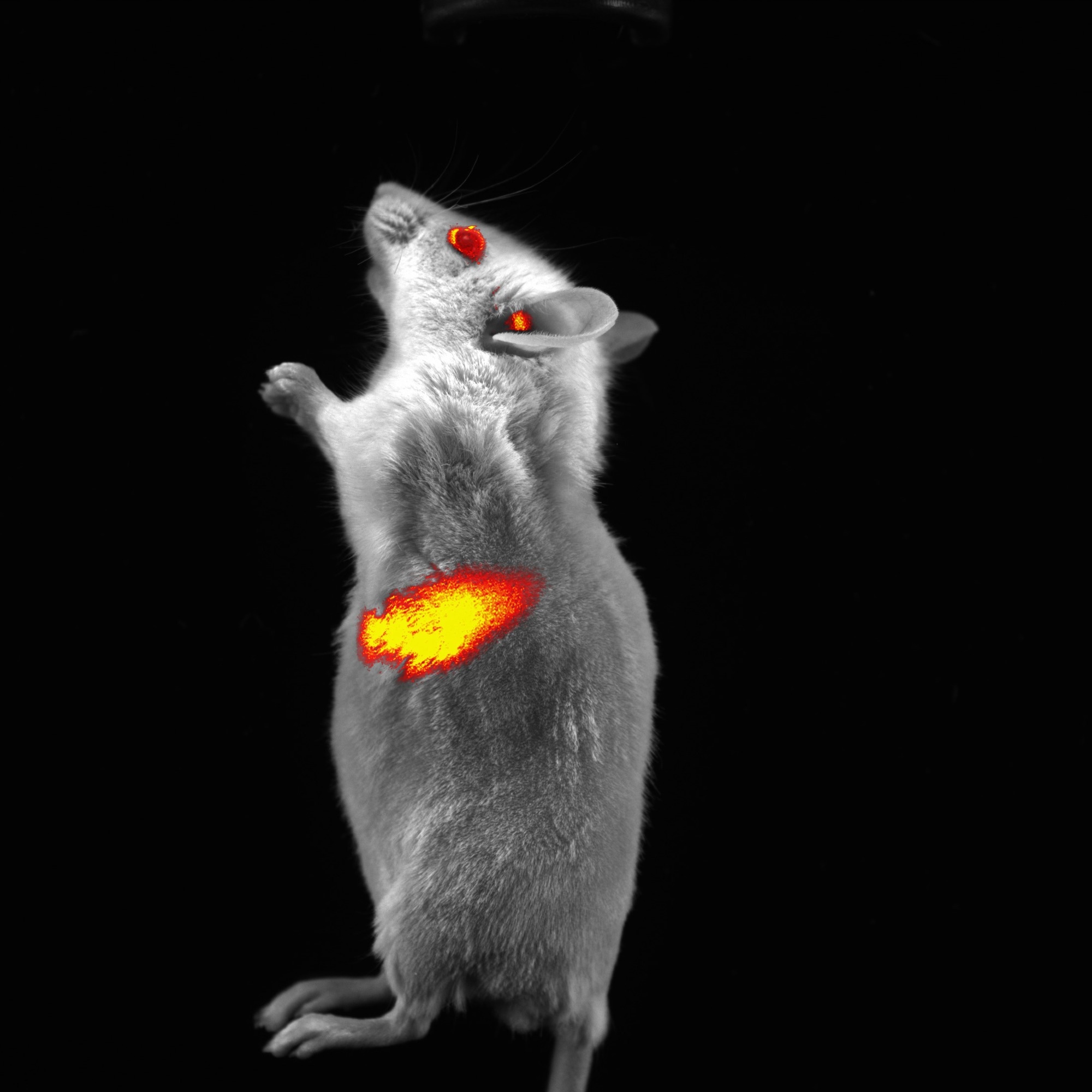
Image Credit: Scintica Instrumentation Inc.
How can NIR-II imaging support longitudinal studies in which researchers need to monitor biological processes repeatedly and noninvasively over time?
NIR-II supports longitudinal studies because it is non-invasive, non-ionizing, and efficient. By affording access to light emitted from deep tissues, researchers no longer need to wait until a terminal endpoint to evaluate organs ex vivo. Instead, biodistribution can be continuously monitored in vivo using repeated measures.
Animals can therefore serve as their own controls at baseline, reducing intra-animal variability, strengthening the study’s statistical power, and requiring fewer animals overall. By refining our animal testing methods, the Newton FT-900 supports the three Rs of research (replacement, reduction, and refinement).
What types of probes, reporters, or experimental models are helping drive the adoption of NIR-II imaging in preclinical research?
There is an increasing need for commercially available NIR-II fluorophores. The Newton FT-900 facilitates proper evaluation of probe radiance, target specificity, and metabolism/clearance. This includes small molecule dyes, semiconductor quantum dots, down conversion rare-earth nanoparticles, carbon dots, single-walled carbon nanotubes, and aggregation-induced emission (AIE) probes.
What common misconceptions do researchers have about NIR-II imaging, especially around complexity, cost, workflow integration, or comparison with more established modalities?
Researchers may assume that, as an all-in-one platform, the Newton FT-900 must be complex and expensive. Instead, the system offers a user-friendly interface that makes it easy to learn and engage with, plenty of automation with pre-built applications to choose from, and a cost-effective price that is justifiable in your next grant application.
Other platforms lack the integration of BLI, NIR-I, and NIR-II, requiring researchers to purchase two separate systems to achieve what the Newton FT-900 can do in a single device. This is especially true when compared to other expensive technologies, such as MRI, PET, and SPECT. To install a Newton FT-900 in the lab, no special room modifications or changes to the building infrastructure are necessary, helping to keep costs low.
From an implementation standpoint, what should labs consider before adding NIR-II imaging to their workflow?
One consideration could be the lab’s probe strategy: they may be planning to develop novel dyes or use common fluorophores with longtail emissions into the NIR-II range, or maybe they want to implement the Newton FT900 to future-proof their lab for when more NIR-II dyes become commercially available.
Collectively, many labs prefer a unified platform such as the Newton FT-900, which facilitates the adoption of NIR-II capabilities without disrupting established BLI and FLI studies. Taken together, this minimizes training time while preserving comparability and interpretability with historical data.
How does the Newton FT-900 address the need for flexibility across VIS, NIR-I, NIR-II/SWIR, fluorescence, and bioluminescence imaging in a single platform?
The Newton FT-900 boasts a dual camera architecture to deliver the most complete and versatile in vivo optical imaging system. The DARQ-11 scientific-grade 16-bit CCD camera is used for 2D and 3D BLI, and 2D FLI in the VIS and NIR-I ranges (400–900 nm). Using the InGaAs SWIR camera, 2D FLI in the NIR-II range (900–1700 nm) can be performed.

Image Credit: Scintica Instrumentation Inc.
One of the key features of the Newton FT-900 is its dual-camera architecture. What performance advantages does this provide for researchers working across multiple optical imaging modalities?
With two CCD detectors, researchers can acquire an image with the VIS camera (400–900 nm), followed by a sequential acquisition using the SWIR/NIR-II camera (900–1700 nm). As an example, drug delivery of a tumor-specific NIR-II nanoparticle can be visualized for a deep luciferase-expressing tumor. Therein, images can be captured sequentially and merged to demonstrate the full picture.
Looking ahead, how do you see NIR-II imaging changing preclinical research workflows, and what role could systems like the Newton FT-900 play in making this technology more accessible to research teams?
Technologies that leverage NIR-II optical imaging, such as the Newton FT-900, will be key for improving translational research. The implementation of this equipment will not disrupt workflows but will instead transform the impact of the data it generates.
For context, the foundation of strong translational research is rooted in high-quality preclinical data that more accurately predicts clinical outcomes. At the preclinical studies stage, in vivo animal research is performed to evaluate drug safety, toxicity, PK/PD, and efficacy, while generating insights into disease mechanisms. The results from these studies inform decisions about advancing therapies into humans.
In the context of adopting NIR-II capabilities, researchers can characterize biodistribution and therapeutic responses in real time with better accuracy and precision, thus leading to more translational success.
About Jade Marrow 
Dr. Jade Marrow is a Senior Product Manager at Scintica, where she leads the optical imaging and X-ray portfolios for preclinical research. Her work focuses on translating advanced imaging technologies into practical, high-impact tools for life science researchers. She holds a PhD in Human Health and Nutritional Sciences from the University of Guelph and brings over a decade of research experience spanning human kinetics, cardiovascular physiology, and molecular biology.
About Scintica Instrumentation Inc.
At Scintica, we advance science and medicine by supplying researchers with reliable research instrumentation and equipment. Our carefully selected portfolio of imaging systems, research tools, and supporting technologies is designed to reduce complexity and help scientists focus on what matters most, generating
meaningful results.
We partner closely with the preclinical research community to connect teams with solutions that are scientifically robust and built to support research challenges. From system selection through long-term support, our goal is to make research more productive, efficient, and impactful.
Sponsored Content Policy: News-Medical.net publishes articles and related content that may be derived from sources where we have existing commercial relationships, provided such content adds value to the core editorial ethos of News-Medical.net, which is to educate and inform site visitors interested in medical research, science, medical devices and treatments.