The versatile Newton 7.0 is an advanced optical imaging system that can conduct bioluminescence, fluorescence, and 3D tomographic imaging - all in a single device. It is perfect for in vitro, ex vivo, and in vivo imaging applications as well as simultaneous imaging of several specimens due to its sophisticated features and easy-to-use interface.
Key to the system is a cutting-edge 4.6 megapixel CCD camera with one of the biggest apertures on the market. For a range of luciferase enzymes and fluorophores frequently employed in preclinical research, this camera offers exceptional sensitivity, enabling quick and effective signal capture. Longitudinal studies can save significant time, thanks to the user-friendly tools and straightforward workflow suited for multiple users.
What Makes the NEWTON Optical Systems Stand Out?
Video Credit: Scintica Instrumentation Inc./YouTube.com
Features and benefits
The Newton 7.0 is an inventive optical bioluminescence, fluorescence, and 3D tomographic imaging system developed with the user in mind.
State-of-the-art camera technology
- Scientific grade 0 16-bit CCD
- 4.6 MP Native Resolution
- 10 MP Image Resolution
- -90 ºC Absolute Cooling
- f/0.7 Lens Aperture
- 4.8 Optical Density
Powerful fluorescent excitation
Eight excitation channels, covering the visible and near-infrared spectra, are included in the Newton 7.0. The illuminating light is firmly controlled by two strong Laser Class II arrays, producing a direct, intense light.
Motorized darkroom with adjustable field-of-view
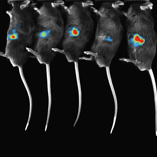
The camera (Z-axis) and animal pad (X/Y axis) can be fully motorized to move across both the macro imaging FOV (6 × 6 cm) and the full FOV (20 × 20 cm). Up to five mice can be imaged at a time, enabled by Vilber’s innovative darkroom archictecture.
Full spectrum tunability
Eight excitation channels and eight emission filters, covering the whole spectrum from blue to infrared, make it powerful.
All of the most widely used fluorophores can be seamlessly imaged by using hard-coated narrow bandpass filters to gather emission wavelengths and minimize signal cross talk.
3D optical tomography
Bioluminescent signals can be reconstructed in three dimensions and overlaid onto topographical models of the imaging subject using an integrated 3D tomography module.
To better understand anatomical and deeper tissue features, the mouse organs and bones can be superimposed onto the topographical model using the digital organ and bone library.
License-free acquisition and analysis software
Free updates and unlimited licenses are included with the user-friendly software.
Both novice and expert users can easily image their subjects with either custom acquisition methods or the factory preset acquisition protocols.
Data that is fully consistent with GLP and CFR21 can be exported in 16-bit.tiff or 8-bit.jpg formats.
Save ROI and analysis templates in the analysis module to analyze up to ten photos side by side at high throughput.
Imaging modes
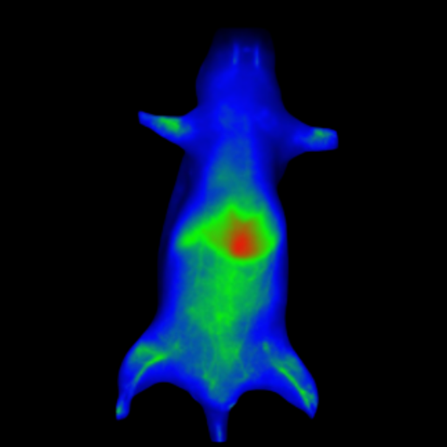
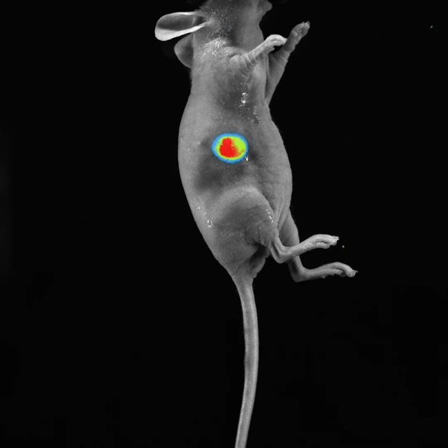
Fluorescence imaging
Vilber's dynamic range of emission filters can be employed in fluorescence imaging to identify fluorescent reporter genes or dyes in vivo.
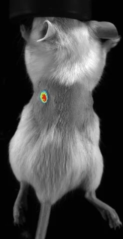
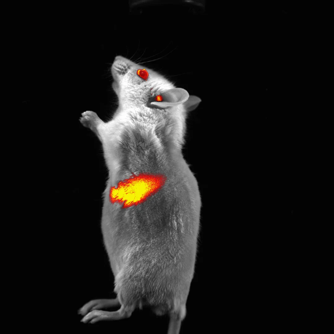
Subcutaneous tumor expressing mCherry. Image Credit: Scintica Instrumentation Inc.
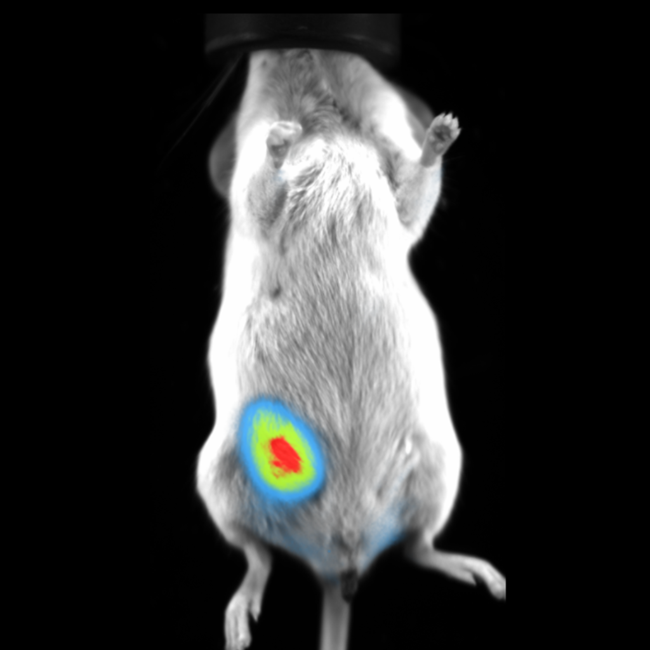
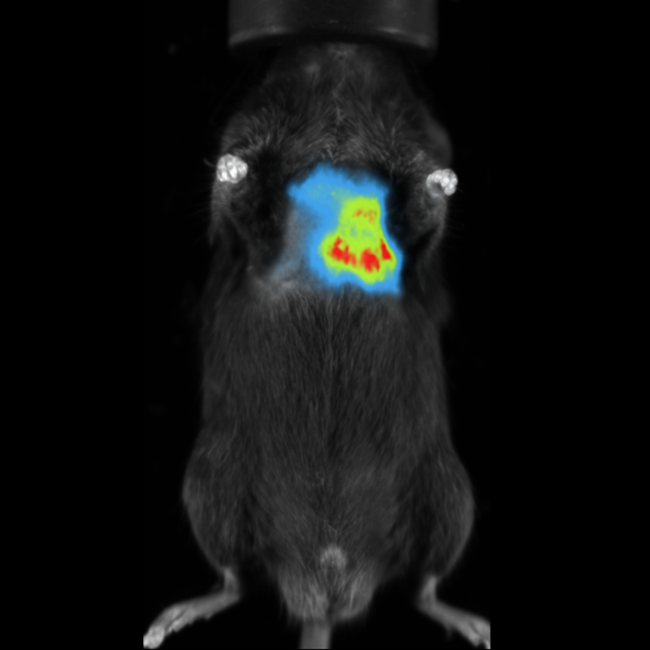
Bioluminescence imaging
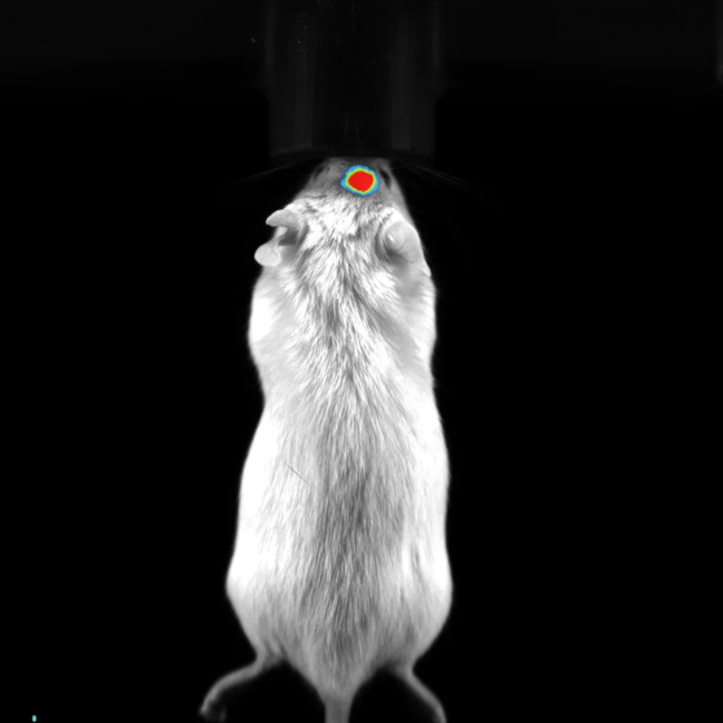
Bioluminescence imaging accurately detects luciferase-expressing or secreting molecules in the target tissue.
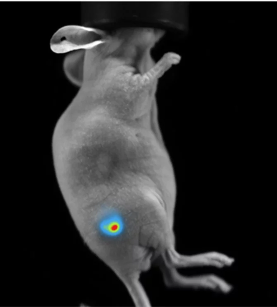
Subcutaneous tumor expressing firefly luciferase. Image Credit: Scintica Instrumentation Inc.
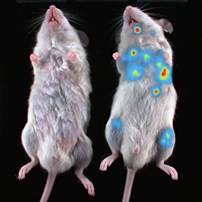
Multispectral in vivo imaging
Different fluorescent dyes or luciferase enzyme/substrate combinations can be used to provide multispectral in vivo imaging. The same image can have up to three signals superimposed on it.
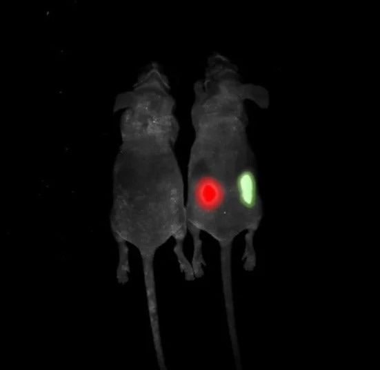
Multispectral imaging. Image Credit: Scintica Instrumentation Inc.
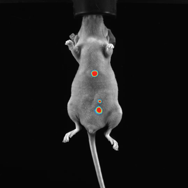
Longitudinal imaging
- A longitudinal image sequence can be created by arranging images that were taken at various times. For instance, pictures taken on various days after an experimental therapy could be used to create a time series
- The image data is then compared by the software over the course of the experimental treatment

Image Credit: Scintica Instrumentation Inc.
Models and specifications
NEWTON 7.0
MODELS BT100 and BT500
- Bioluminescence Detection
- In vivo / in vitro optical imaging platform
- 3D BLI Optical Tomography
- Mice and Rats
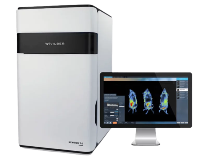
Image Credit: Scintica Instrumentation Inc.
NEWTON 7.0
MODELS FT100 and FT500
- Bioluminescence Detection
- VIS and NIR-1 Fluorescence
- In vivo / in vitro optical imaging platform
- 3D BLI Optical Tomography
- Mice and Rats
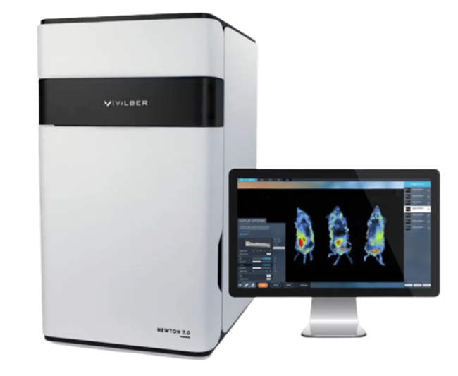
Image Credit: Scintica Instrumentation Inc.
Newton 7.0 Bio
Plant Imaging
- Fluorescence Detection Picogram
- In vivo / in vitro optical imaging platform
- Bioluminescence Detection Fentogram
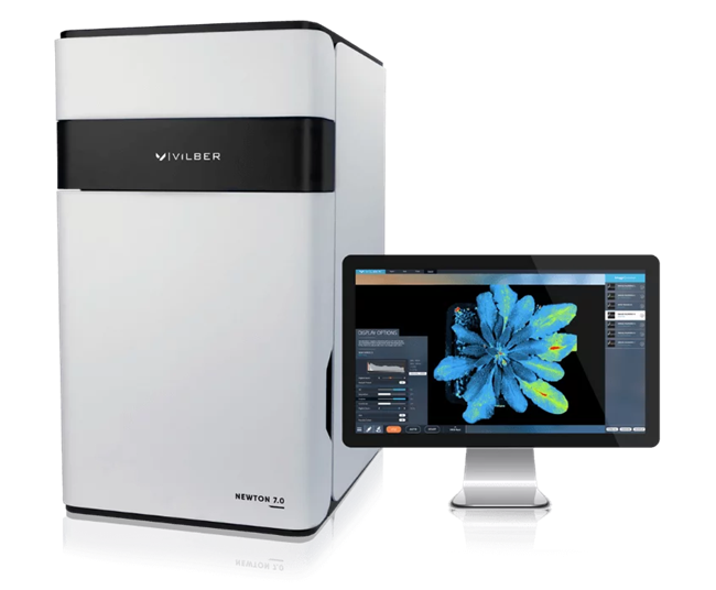
Image Credit: Scintica Instrumentation Inc.
Camera
All models
- Cooling: -120 °C Delta
- 16-bit Scientific Grade CCD Camera
- Cooling: -90 °C absolute
Lens
All models
- Focal Length Motorized lens
- Proprietary V.070 – Fixed
- Aperture: f/0.7
Resolution
All models
- Resolution: 2160 × 2160
- Monochrome and Color Imaging
Emission
All models
- Eight Narrow Band-pass filters included as standard 500 / 550 / 600 / 650/ 700/ 750 / 800 / 850 nm
- 11-Position motorized filter wheel for all models
Excitation
Source: Scintica Instrumentation Inc.
| |
BT100 |
BT500 |
FT100 |
FT500 |
BIO |
| White-Light |
Dual EPI-
White Light
LED Panels |
Dual EPI-
White Light
LED Panels |
Dual EPI-
White Light
LED Panels |
Dual EPI-
White Light
LED Panels |
Dual EPI-
White Light
LED Panels |
|
| Fluorescence |
Upgradeable
to
Fluorescence |
Upgradeable
to
Fluorescence |
8 Fluorescent
Channels Included
440 / 480 / 540 /
580 / 640 / 680 /
740 / 780 nm |
8 Fluorescent
Channels Included
440 / 480 / 540 /
580 / 640 / 680 /
740 / 780 nm |
8 Fluorescent
Channels Included
440 / 480 / 540 /
580 / 640 / 680 /
740 / 780 nm |
Emission
Source: Scintica Instrumentation Inc.
| |
BT100 |
BT500 |
FT100 |
FT500 |
BIO |
Filter
Wheel |
11-position Motorized Filter Wheel |
11-position Motorized
Filter Wheel |
11-position Motorized Filter Wheel |
11-position Motorized Filter Wheel |
11-position Motorized Filter Wheel |
|
Emission
Filters |
4 Narrow Band-pass filters included for BLI Tomography:
500/550/600/
650 nm |
4 Narrow Band-pass filters included for BLI Tomography: 500/550/600/
650 nm |
8 Narrow Band-pass filters included:
500/550/600/
650/700/750/
800/850 nm |
8 Narrow Band-pass filters included:
500/550/600/
650/700/750/
800/850 nm |
8 Narrow Band-pass filters included:
500/550/600/
650/
700/750/
800/850 nm |
Darkroom
Source: Scintica Instrumentation Inc.
| |
BT100 |
BT500 |
FT100 |
FT500 |
BIO |
| Motorization |
- Fixed Camera
- Fixed Animal Stage
|
- Z-Axis Motorized Camera
- X/Y-Axis Motorized Animal Stage
|
- Fixed Camera
- Fixed Animal Stage
|
- Z-Axis Motorized Camera
- X/Y-Axis Motorized Animal Stage
|
- Z-axis Motorized Camera
- 15 ° Tilting Sample Stage
|
|
| Animal Handling |
- Heated Mouse Bed (+37 °C) included
- Animal breathers included
|
- Heated Mouse Bed (+37 °C) included
- Animal breathers included
|
- Heated Mouse Bed (+37 °C) included
- Animal breathers included
|
- Heated Mouse Bed (+37 °C) included
- Animal breathers included
|
|
Animal handling - all models
- Animal breathers (included)
- Heated Mouse Bed (+37 °C) (included)
(Not Included with Newton 7.0 BIO)
Accessories / Add-ons
Source: Scintica Instrumentation Inc.
| |
BT100 |
BT500 |
FT100 |
FT500 |
BIO |
| Monitorization |
Fixed
Camera
Fixed
Animal
Stage |
Z-Axis
Motorized
Camera
X/Y-Axis
Motorized
Animal Stage |
Fixed
Camera
Fixed
Animal
Stage |
Z-Axis
Motorized
Camera
X/Y-Axis
Motorized
Animal Stage |
Z-axis
Motorized
Camera
15 ° Tilting
Sample
Stage |
Applications
Oncology
In preclinical animal models, optical imaging can be used to non-invasively track the development and dissemination of cancer throughout the body.
Immunology
Monitoring different immune cell types can significantly increase knowledge of their physiology and the creation of novel treatment approaches.
Infectious disease
Monitoring different immune cell types can greatly advance the knowledge of their physiology and the creation of novel treatment approaches.
Neurology
Optical imaging can be used to evaluate new targeted treatments in the brain and spinal cord, as well as track the development of different neurodegenerative disorders.
Biodistribution studies
Optical imaging has a unique advantage in preclinical biodistribution studies as it can image the entire subject - a single image can provide measurements for several organs throughout the body.
Newton 7.0 image gallery
BLI Imaging of Orthotropic Mammary Fat Pad Tumor in Mouse. Image Credit: Scintica Instrumentation Inc.
Fluorescent Nanoprimer Distribution After IV Injection in Mouse. Image Credit: Scintica Instrumentation Inc.
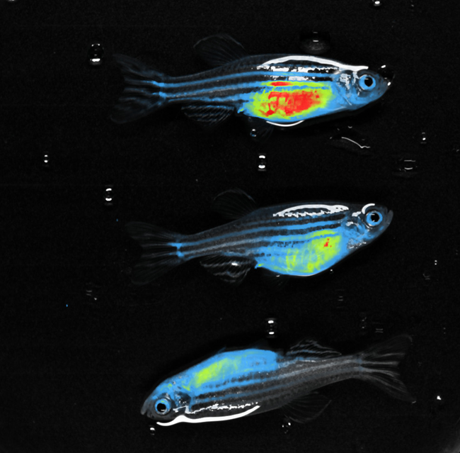
RED FLI - Zebrafish. Image Credit: Scintica Instrumentation Inc.

Plants- TUYV GFP Pot. Image Credit: Scintica Instrumentation Inc.
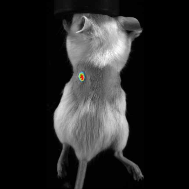
mCherry Fluorescence Imaging in Subcutaneous Tumor in Mouse. Image Credit: Scintica Instrumentation Inc.
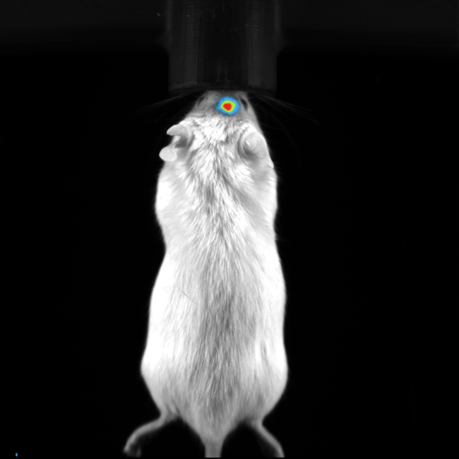
BLI Image of Orthotropic Brain Tumor in Mouse. Image Credit: Scintica Instrumentation Inc.
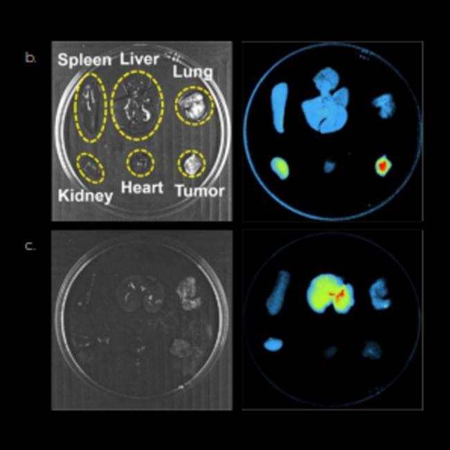
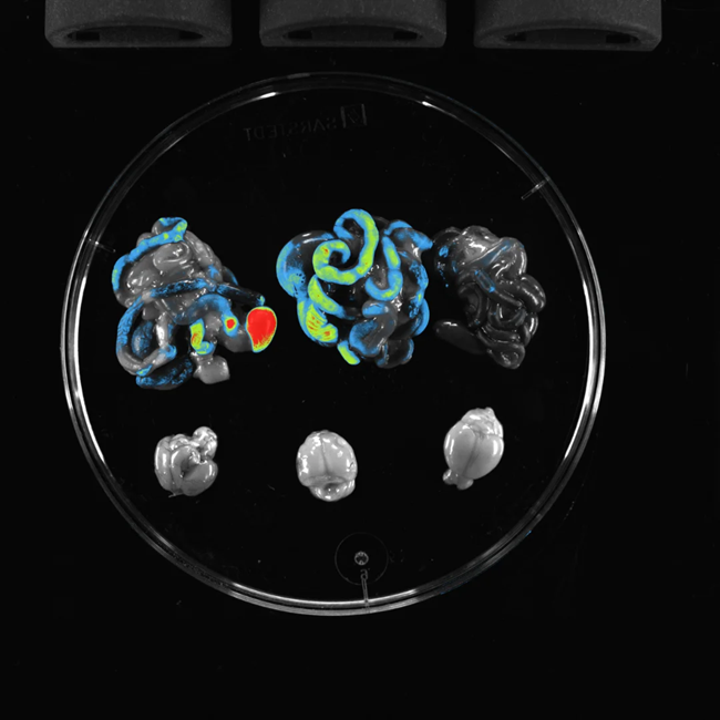
Spleen, Liver, Lung, Kidney, Heart, Tumor. Image Credit: Scintica Instrumentation Inc.
BLI Image of Orthotropic Lung Metastases in Mouse. Image Credit: Scintica Instrumentation Inc.
BIOLUMINESCENCE - Color BF - LIH Luxembourg. Image Credit: Scintica Instrumentation Inc.
BIOLUMINESCENCE - Luc Tumor - Courtesy of CEA Orsay (France). Image Credit: Scintica Instrumentation Inc.
BIOLUMINESCENCE - Tumor and Metastasis - Courtesy of Warsaw University (Poland). Image Credit: Scintica Instrumentation Inc.
BIOLUMINESCENCE - Tumors - Courtesy of Lausanne Uni (Switzerland)_ana. Image Credit: Scintica Instrumentation Inc.
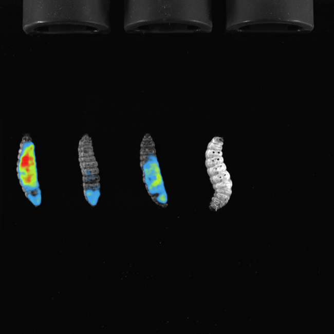
BIOLUMINESCENCE - zWorms - Innsbruck. Image Credit: Scintica Instrumentation Inc.
FLUORESCENCE - 780 nm NIR Liver Infection - Courtesy of Polatom (Poland) (1). Image Credit: Scintica Instrumentation Inc.
Long-Term Optical Imaging Reveals Bone Marrow Clearance and Extramedullary Escape in BCMA CAR-NK Therapy. Image Credit: Scintica Instrumentation Inc.
ZEX-VIVO - GFP Organs. Image Credit: Scintica Instrumentation Inc.