A cutting-edge benchtop micro-CT platform, the inCiTe™ 3D X-ray Microscope provides high-resolution, three-dimensional vision of biological tissues, bone microarchitecture, and cutting-edge materials.
Its phase contrast imaging characteristics make it possible to see minute structural details in low-density specimens, implant-tissue interfaces, and trabecular and cortical bone that are challenging to see with traditional X-ray methods. The inCiTe™ 3D is a powerful yet small device that revolutionizes preclinical orthopedic research applications by providing remarkable insights.
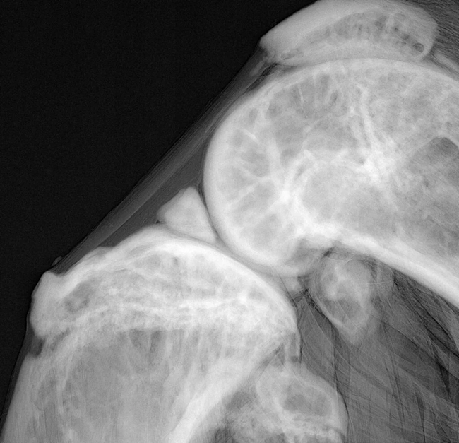
Image Credit: Scintica Instrumentation Inc.
Features and benefits
- Propagation-based X-Ray Phase Contrast
- Ex vivo Imaging of Biological Samples
- User Friendly Interface
- Streamlined Workflow
- Ultra High Resolution
- Quicker Scan Time
Models and specifications
inCiTe™ 3D X-ray microscope 5 μm
Source specifications
- Smallest Spot Size: 5 μm (at 4 W)
- Type: Maintenance-free, sealed
- Maximum Power: 20 W
- Voltage: 40-100 kV
inCiTe™ 3D X-ray microscope 2 μm
Source specifications
- Smallest Spot Size: 2 μm (at 4 W)
- Type: Maintenance-free, sealed
- Maximum Power: 16 W
- Voltage: 40-100 kV
Source: Scintica Instrumentation Inc.
| General Specifications |
inCiTe™ 3D X-ray Microscope 5 μm / 2 μm |
| Maximum sample/scanning diameter |
25 mm |
| Maximum scanning length |
30 mm |
| Maximum sample length |
100 mm |
| Image format |
Raw (no header), 16-bit unsigned, little endian |
| Spatial resolution |
11 μm (0.5 MTF at 45 cycles/mm)
5.6 μm (0.1 MTF at 90 cycles/mm) |
| X-Ray phase contrast |
Propagation-based |
| Source-to-object distance |
44-410 mm |
| Source-to-detector distance |
84-450 mm |
| Lateral object translation |
50 mm |
| Vertical object translation |
12.7 mm |
| Rotational stepping |
0.01 degrees (min.) |
| Detector type |
Selenium-CMOS direct conversion |
| Detector format |
16 MP (4k x 4k pixels) |
| Detector area |
32 × 32 mm2 |
| Pixel pitch |
8 μm |
| Pixel size at maximum magnification |
0.8 μm |
| Radiation safety |
< 4 μSv/hr at 5 cm distance from any accessible surface |
| Dimensions (D x W x H) |
48 cm (D) x 150 cm (W) x 60 cm (H) |
| Weight |
280 kg |
| Installation requirements |
- 120V AC power
- 10-30 °C temperature
- < 85 % humidity
- Zero condensation
|
Applications
- Post-injury Fracture Healing
- Musculoskeletal Disorders
- Preclinical Imaging
- Bone Diseases
- Orthopedics
Imaging gallery
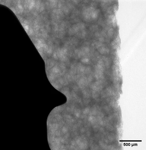
Image Credit: Scintica Instrumentation Inc.
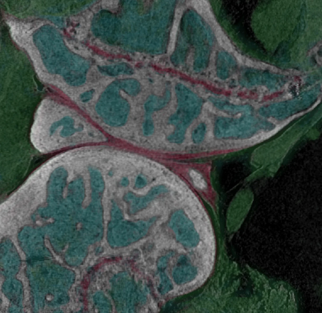
Image Credit: Scintica Instrumentation Inc.
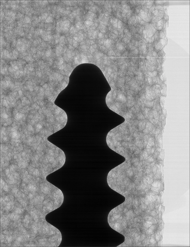
Image Credit: Scintica Instrumentation Inc.
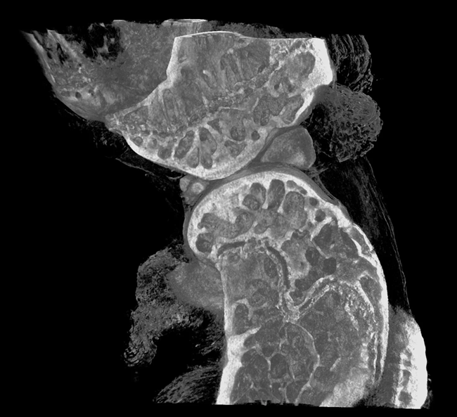
Image Credit: Scintica Instrumentation Inc.