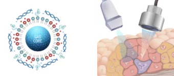
The USphere Series of microbubble contrast agents has been carefully designed for preclinical imaging, operating efficiently at frequencies ranging from 1 to 40 MHz. Their small size distribution (1.1-1.4 µm) and high concentration (∼2.5×10¹⁰ bubbles/ml) render them exceptionally suitable for application across various species, including mice, rats, rabbits, and non-human primates.
A diverse array of agents are available: for imaging perfusion, targeting specific biomarkers, as well as agents containing fluorescent dyes for multi-modal imaging, and those intended for targeted drug delivery or gene transfection - among others.
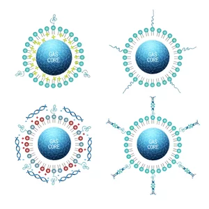
Image Credit: Scintica Instrumentation Inc
The USphere Series of microbubble contrast agents has been meticulously crafted for application across various species used in preclinical research, including mice, rats, rabbits, and even pigs; these agents have been rigorously tested and demonstrated effective performance at frequencies ranging from 1 to 40 MHz.
In particular, these agents have undergone testing on the Prospect T1 high-frequency ultrasound system, where they have shown commendable performance in both linear and harmonic contrast imaging modes.
A diverse array of bubble configurations is available, some designed specifically for studying perfusion, others to assist in biomarker detection, and yet others for facilitating ultrasound-mediated drug or gene delivery, along with more sophisticated theranostic applications.
The critical factors influencing the particle size of the microbubble have been determined; through precise adjustments to the lipid formulation, the USphere agents have been engineered to achieve the smallest size distribution (1.1-1.4 µm) compared to competitors in the current market.
The USphere agents feature a phospholipid shell filled with perfluoropropane (C3F8) or, optionally, perfluorobutane (C4F10). The concentration of microbubbles has been optimized for small animal imaging (∼2.5×10¹⁰ bubbles/ml), ensuring that low volume injections yield sufficient contrast signals in vivo without overburdening their circulatory systems.
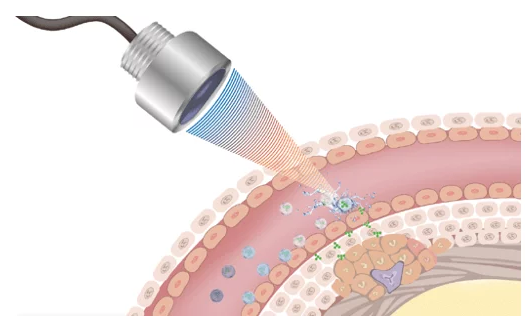
Image Credit: Scintica Instrumentation Inc.
Microbubble contrast agents are capable of visualizing all levels of vasculature, offering detailed perfusion information that may be pertinent in tumor and abdominal organ imaging, as well as in drug development studies, to name just a few.
Features and benefits
High concentration of microbubbles per ml of activated contrast agent (∼1-5×10¹⁰ bubbles/ml)
When handling small animals like mice or rats, the recommended injection volume (1-20 µL) is minimal enough to avoid overloading the circulatory system, while still providing an adequate amount of microbubbles to achieve the intended contrast enhancement.
Additionally, further dilution of the microbubbles can be performed using sterile saline to facilitate more precise delivery of limited quantities of microbubbles.
Small size distribution (1.1-1.4 µm) of activated microbubbles
To enable precise perfusion properties of the microvasculature, the microbubbles must be nanoscale in size.
Once activated, the microbubble concentration is stable for 72 hours when stored at 2-8 ℃
The microbubbles' stability for a duration of three days enables the user to conduct the necessary studies within that timeframe, making full use of the entire volume of the agent and preventing the wastage of any excess agent that cannot be utilized.
Microbubbles have been shown to work at a wide range of frequencies (1-40 MHz)
The microbubbles are applicable to a diverse range of species, including mice, rats, rabbits, non-human primates, and pigs.
The microbubbles have been utilized across a broad spectrum of ultrasound systems from different manufacturers, employing both linear and non-linear/harmonic contrast imaging modes.
Models and specifications
Source: Scintica Instrumentation Inc.
| Product Line |
Product Name |
Size Distribution |
Concentration |
| USphere™ Prime |
Prime C3F8 |
1.1-1.4 µm |
> 2.5x10 microbubbles/ml;
Each vial contains 0.8 ml |
| USphere™ Prime |
Prime C4F10 |
1.1-1.4 µm |
> 2.5x10 microbubbles/ml;
Each vial contains 0.8 ml |
| USphere™ Tracer |
Tracer FD-Red – C3F8 |
1.1-1.4 µm |
> 2.5x10 microbubbles/ml;
Each vial contains 0.8 ml |
| USphere™ Tracer |
Tracer FD-Green – C3F8 |
1.1-1.4 µm |
> 2.5x10 microbubbles/ml;
Each vial contains 0.8 ml |
| USphere™ Labeler |
Labeler LB (Biotin) – C3F8 |
1.1-1.4 µm |
> 2.5x10 microbubbles/ml;
Each vial contains 0.8 ml |
| USphere™ Labeler |
Labeler LA (Biotin and
Avidin) – C3F8 |
1.1-1.4 µm |
> 2.5x10 microbubbles/ml;
Each vial contains 0.8 ml |
| USphere™ Labeler |
Labeler FLA (LA with
Fluorescence) – C3F8 |
1.1-1.4 µm |
> 2.5x10 microbubbles/ml;
Each vial contains 0.8 ml |
| USphere™ Labeler |
Labeler LS (Biotin and
Streptavidin) – C3F8 |
1.1-1.4 µm |
> 2.5x10 microbubbles/ml;
Each vial contains 0.8 ml |
| USphere™Trans+ |
Trans+ TP – C3F8 |
1.1-1.4 µm |
> 2.5x10 microbubbles/ml;
Each vial contains 0.8 ml |
| USphere™ Tracer |
Tracer FD-Red – C4F10 |
1.1-1.4 µm |
> 2.5x10 microbubbles/ml;
Each vial contains 0.8 ml |
| USpher™ Tracer |
Tracer FD-Green – C4F10 |
1.1-1.4 µm |
> 2.5x10 microbubbles/ml;
Each vial contains 0.8 ml |
| USpher™ Labeler |
Labeler LB (Biotin) – C4F10 |
1.1-1.4 µm |
> 2.5x10 microbubbles/ml;
Each vial contains 0.8 ml |
| USpher™ Labeler |
Labeler LA (Biotin and
Avidin) – CF410 |
1.1-1.4 µm |
> 2.5x10 microbubbles/ml;
Each vial contains 0.8 ml |
| USphere™ Labeler |
Labeler FLA (LA with
Fluorescence) – C4F10 |
1.1-1.4 µm |
> 2.5x10 microbubbles/ml;
Each vial contains 0.8 ml |
| USphere™ Labeler |
Labeler LS (Biotin and
Streptavidin) – C4F10 |
1.1-1.4 µm |
> 2.5x10 microbubbles/ml;
Each vial contains 0.8 ml |
| USphere™Trans+ |
Trans+ TP – C4F10 |
1.1-1.4 µm |
> 2.5x10 microbubbles/ml;
Each vial contains 0. 8ml |
Applications
Perfusion imaging
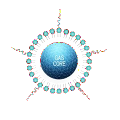
USphere Prime. Image Credit: Scintica Instrumentation Inc.
USphere Prime agents consist of gas-filled microbubbles (perfluoropropane or perfluorobutane) encased in a phospholipid shell. The small size distribution enables these microbubbles to navigate effortlessly through all tiers of the vascular system, including the capillaries.
Targeted molecular imaging
USphere Labeler agents have been functionalized with various moieties, including biotin, avidin, and/or streptavidin, enabling the incorporation of specific targeting agents like small molecules and antibodies. These agents can be tailored to target particular biomarkers of interest.
Multi-modal imaging
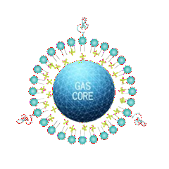
USphere Tracer. Image Credit: Scintica Instrumentation Inc.
The USphere Tracer agents, along with the USphere Labeler featuring Fluorescence, have been enhanced to incorporate a fluorescent dye, enabling multi-modal imaging with a fluorescence imaging system.
This enhancement can be significant in pharmacodynamics/pharmacokinetics or other scenarios where fluorescent imaging plays a crucial role.
Drug delivery and gene transfection
Usphere Trans+. Image Credit: Scintica Instrumentation Inc.
The USphere Trans+ agents have been modified to possess a positively charged surface. These microbubbles are capable of delivering a substantial payload, thereby enhancing the targeting, effectiveness, and simplicity of drug delivery and gene transfection compared to conventional methods.