The VILBER Fusion systems are designed for quantification-grade imaging.
Producing quantitative and repeatable statistics from chemiluminescent western blot data presents unique problems. Low dynamic range of detection and challenges in precisely identifying the limit of detection are the main causes of these problems.
Fusion systems eliminate these concerns with High Sensitivity Reading (HSR) camera technology, which provides consistent dynamic range, linearity, and unrivaled sensitivity for the lowest limit of detection. By lowering noise, the sophisticated camera technology makes it possible for weak signals to be distinguished from the backdrop.
Fusion systems can produce consistent and reproducible data regardless of the chemiluminescence time course. The intensity/time profile of chemiluminescence shows a spike at first, followed by a protracted emission at a pseudo-plateau level and a fall. VILBER Fusion Automatic imaging mode maintains the largest possible image dynamic while compensating for the chemiluminescence reaction's time course through exposure time adjustments.
VILBER Fusion Fluorescence and Chemiluminescence systems are excellent multimodal imaging solutions for molecular biology research. They span the range of applications for chemiluminescence, multiplexed fluorescence, and chemifluorescence. In all DNA, RNA, and protein-based applications, it provides the highest sensitivity, speed, and lowest detection limits. The technology used by the Fusion FX7 to capture images is cutting edge, as is the ease with which users may process and analyze them. The Fusion system is suited for publication, quantification, and documentation-grade imaging.
Leading life science firm VILBER creates and produces fluorescence and chemiluminescence imaging devices for anything from cell biology research to small animal applications. Vilber, a pioneer in the field of molecular imaging, was established in 1954 and has equipped over 20,000 laboratories globally. Their products are used daily by an estimated 60,000 researchers in more than 100 countries.
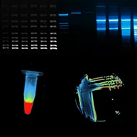
Image Credit: Scintica Instrumentation Inc.
Features and benefits
The VILBER Fusion line of systems is ideal for quantification-grade imaging. Chemiluminescent western blot data pose distinct challenges in producing quantifiable and reproducible data. The low dynamic range of detection and the challenge of precisely identifying the limit of detection are the main causes of these problems.
Thanks to a High Sensitivity Reading (HSR) camera technology that provides dependable dynamic range, linearity, and unmatched sensitivity at the lowest limit of detection, Fusion systems do away with all these problems. By lowering noise, the sophisticated camera technology makes it possible for weak signals to be distinguished from the background.
Furthermore, regardless of a chemiluminescence time course, the Fusion systems may produce reliable and repeatable data. The intensity/time profile of chemiluminescence shows a spike at first, followed by an extended emission at a pseudo-plateau level and a drop. VILBER Fusions Automatic imaging mode modifies the exposure time to adapt for the chemiluminescence reaction's time course while preserving the maximum image dynamic.
State-of-the-art camera technology
With an extremely low signal to noise ratio, the sophisticated camera and optics offer enhanced sensitivity to either chemiluminescence or fluorescence signals. Samples with extremely low and high signals can be scanned without saturation, thanks to the high optical density, producing quantitative findings.
- 10 megapixel image resolution
- Scientific grade 16-bit CCD
- -90 ºC delta cooling
- 4.8 Optical Density
- f/0.7 aperture
One click to image
The Fusion FX7 is made to be as user-friendly as possible. This system is plug-and-play due to its easy installation and user-friendly interface. The most user-friendly software for imaging is Fusion FX7. After placing the blot on the tray, choosing the application needed, and clicking "Start," the system automatically exposes both the marker and blot images and merges them.
Powerful fluorescence excitation
Seven adjustable excitation channels in the ultraviolet, visible, and deep infrared spectrums are available with the Fusion. A very small excitation filter further restricts the extremely limited LED spectrum.
Free software
Complementary free software for image analysis, editing, and enhancement. Additionally, software upgrades are always free.
Spectral unmixing
When using distinct luciferase enzymes or fluorescent dyes, spectral unmixing can be achieved for both chemiluminescence and fluorescence imaging.
This contains algorithms that eliminate crosstalk between the various signals, enabling a single reporter's signal to be present on each channel.
Complete and practical
The system is made in France using premium components. It is very simple to operate and has a small footprint.
Advanced software
The system is made in France using premium components. It is very simple to operate and has a small footprint.
3D dynamic scan
The software that comes with the gel doc systems gives improved options for image editing and analysis, making it easy to create photographs that are ready for publishing.
Use the quantification, molecular weight calculation, or distance (Rf) calculation modules to analyze images.
Models and specifications
Fusion - Absolute
Combined fluorescence and chemiluminescence for western blot and gel documentation imaging
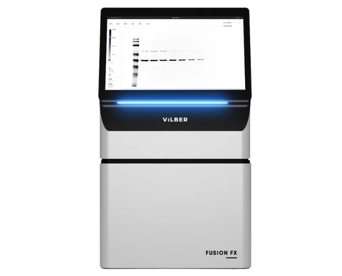
Image Credit: Scintica Instrumentation Inc.
Fusion - FX7
Chemiluminescence and fluorescence
Can be updated at any moment to support a variety of imaging applications, such as standard gel documentation.
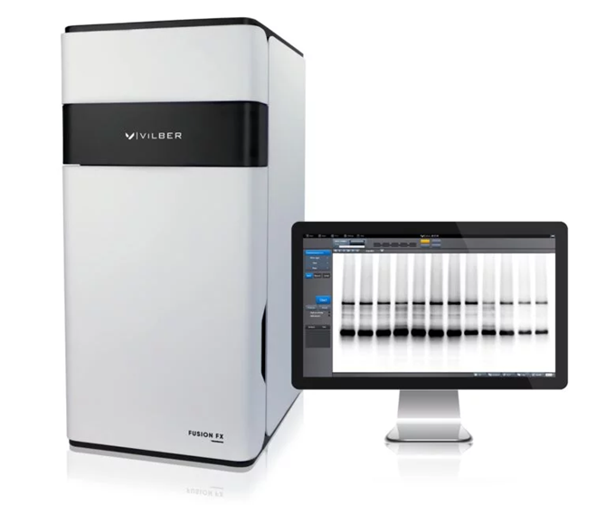
Image Credit: Scintica Instrumentation Inc.
Fusion - FX6
Chemiluminescence and fluorescence
Upgradable at any time to accommodate various imaging applications (e.g., standard gel documentation)
Image Credit: Scintica Instrumentation Inc.
Fusion - Solo 6S
Chemiluminescence and fluorescence
Can be modified at any time to handle additional imaging applications (for example, standard gel documentation).

Image Credit: Scintica Instrumentation Inc.
Fusion - Solo 6X
Chemiluminescence only
Gel Imaging upgrades need to be bought along with the system.
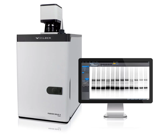
Image Credit: Scintica Instrumentation Inc.
Applications
Chemiluminescence and bioluminescence imaging
- In vivo / in vitro luciferase
- Western, Northern, and Southern Blot
Protein gel imaging and other colometric samples
- Coomassie blue, ponceau S red, silver stain, copper stain
- X-Ray film, SSCOP gels, autorads, colony dishes, colorimetric western blots, flask imaging
Visible RGB and IR/NIR fluorescence imaging
- 2D-DIGE
- Fluorescent Western Blots
- Multiplexing Capabilities (3 colors overlay)
Chemiluminescence and fluorescence western, northern, or Southern blot. Choose the Fusion FX Spectra model according to the applications
- UV: 365 nm
- RGB: 640 nm, 530 nm,-480 nm
- IR/NIR: 780 nm, 640 nm
- 7 Channels: 440 nm, 480 nm, 530 nm, 640 nm, 680 nm, 740 nm, 780 nm
Optional applications include
- Colorimetric stained protein gels, X-Ray film, autorads, SSCP gels, colony dish, and flask imaging with WhiteLight-Pad or UV-Pad + conversion screen
- DNA and RNA gels and fluorescence stain imaging with UV-Pad or Blue-Pad
Optics
Source: Scintica Instrumentation Inc.
| Fusion FX7 |
Rest of Models |
| 16-bit Scientific Grade CCD Camera |
16-bit Scientific Grade CCD Camera |
| Grade 0 / 400-900 nm / 4.8 OD |
Grade 0 / 400-900 nm / 4.8 OD |
| Cooling: -90 °C Delta |
Cooling: -55 °C Delta |
Proprietary V.070 –
Fixed Focal Length Motorized lens |
Proprietary V.070 – Fixed Focal Length Motorized lens |
| Aperture: f/0.7 |
Aperture: f/0.7 |
| Resolution: 10 Megapixels |
Resolution: 20 Megapixels |
| Monochrome & Color imaging |
Monochrome & Color imaging |
Spectra capsules
Source: Scintica Instrumentation Inc.
| Fusion FX7 & FX6 |
Fusion Solo S6 & 6X |
| Capsule Adapter Included |
Capsule Adapter Required |
Excitation epi-illumination
Source: Scintica Instrumentation Inc.
| All Models Except Fusion Solo 6X |
Fusion Solo 6X |
| 7 Customizable channels |
4 Customizable channels |
| Motorized |
Non-Motorized |
| UV, R, G, B, FR, NIR, DIR |
UV, R, G, B, FR, NIR, DIR |
Operating
All models
Dark room
All models
Emission
All models
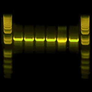
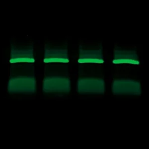
Western Blot imaging gallery
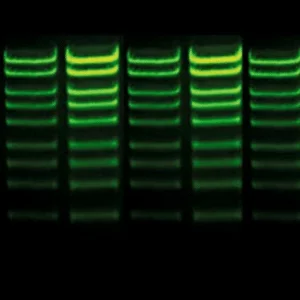
Krypton. Image Credit: Scintica Instrumentation Inc.
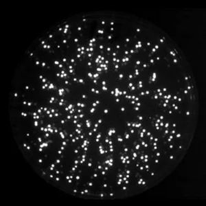
Petri Dish Luciferase. Image Credit: Scintica Instrumentation Inc.
Alexa Fluo- 568. Image Credit: Scintica Instrumentation Inc.
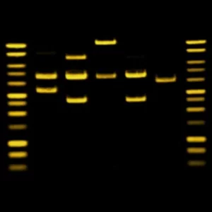
Sybr Gold. Image Credit: Scintica Instrumentation Inc.
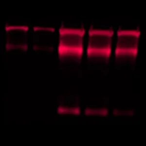
Sypro Ruby. Image Credit: Scintica Instrumentation Inc.
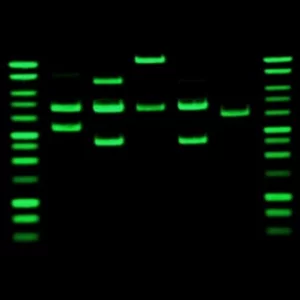
Sybr Green. Image Credit: Scintica Instrumentation Inc.
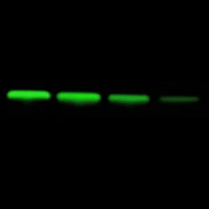
Irdye-800. Image Credit: Scintica Instrumentation Inc.
Qdot-565. Image Credit: Scintica Instrumentation Inc.