Sponsored Content by Binding SiteReviewed by Olivia FrostJul 23 2025
Multiple myeloma (MM) is the second most common hematological malignancy in the United Kingdom (UK), claiming approximately 3000 lives each year, corresponding to at least two percent of all cancer-related deaths.1
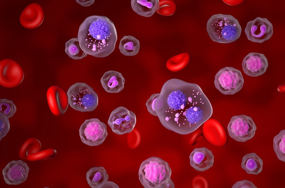
Image Credit: Nemes Laszlo/Shutterstock.com
MM often goes undetected until its later stages because it presents with non-specific symptoms, including fatigue, weight loss, bone pain, and recurrent infections.2,3 However, better awareness, timely diagnosis, and effective management of this challenging condition could help to improve patient outcomes. In fact, early diagnosis has the potential to be the most effective improvement in management available for MM patients, offering them the best chances of a better quality of life and long-term survival.
This article examines the current challenges in diagnosing MM, and how advances in diagnostic technologies - and the systemic changes needed to introduce them - could help patients suffering from this disease.
Understanding multiple myeloma
MM is a malignant cancer of plasma cells in the bone marrow. Plasma cells are a subset of white blood cells responsible for producing antibodies to combat infections, but when these cells become cancerous, they produce large amounts of monoclonal proteins (M proteins) - commonly known as paraproteins - that lack the ability to defend against infections.3 Furthermore, the proliferation of cancerous plasma cells in the bone marrow suppresses the production of healthy blood cells, leading to low counts of red blood cells, other white blood cells, and platelets.3,4
MM can affect bones throughout the body, including the spine, skull, pelvis, and ribs, with symptoms caused by both the growth of the tumour itself (and its effects on the bone structure and the bone marrow microenvironment), and the effect of the monoclonal proteins on other organs. Even advanced MM symptoms are often non-specific and may be mistaken for the effects of aging or other diseases, leading to delayed diagnosis.
Common signs include bone pain (especially in the back or ribs), fatigue, infections, weight loss, and breathlessness.3 As the disease advances, complications such as renal failure, high calcium levels, and bone lesions may develop - summarized by the CRAB criteria: calcium elevation, renal dysfunction, anemia, and bone disease.5
MM typically affects those over 65, though its symptoms and progression can vary.6 Many patients are first diagnosed with monoclonal gammopathy of undetermined significance (MGUS), a precursor condition where plasma cells produce paraproteins without causing organ damage.7 Although MGUS is often discovered incidentally during routine blood tests, it carries a slightly reduced survival rate compared to healthy individuals, with around one percent progressing to MM each year.8 Regular monitoring is vital, as early detection of progression to symptomatic myeloma can prevent organ damage and improve outcomes.

Image Credit: Nemes Laszlo/Shutterstock.com
A heterogenous disease
Excessive paraprotein production is a hallmark of multiple myeloma (MM), but the condition encompasses several distinct subtypes.9 The majority of MM cases (over 80 percent) involve the excessive production of an intact monoclonal immunoglobulin, most frequently of the IgG class, followed by IgA and then IgM.
Alongside these subtypes, rarer forms marked by the overproduction of less common immunoglobulins such as IgD and IgE myelomas exist.10 These rare variants are often more aggressive than IgG and IgA types, requiring specific diagnostic strategies to identify and manage the disease effectively.11,12 In contrast, non-secretory myeloma - a rare subtype affecting one to two percent of patients - is characterized by the production of little to no detectable paraproteins by traditional techniques.13
In addition to intact monoclonal immunoglobulins, many myeloma subtypes also produce other paraproteins, such as free light chains of the kappa (κ) or lambda (λ) types. In cases of light chain-only myeloma - which accounts for over 15% of patients - the disease is defined by the exclusive overproduction of these free light chains. When excreted in urine, they are commonly known as Bence Jones proteins. The presence of serum free light chains (sFLC) is clinically significant and often associated with renal complications, including cast nephropathy and AL amyloidosis. Given the wide variability in myeloma subtypes, a tailored diagnostic and treatment strategy is essential for effective management.
The importance of early diagnosis
The treatment landscape for MM has undergone a significant transformation over the last two decades, driven by a deeper understanding of its molecular biology. However, early diagnosis remains a crucial element in optimal patient management. Advances in MM treatment, including the development of therapies such as proteasome inhibitors, immunomodulators and monoclonal antibodies, have dramatically extended median survival times from around three years to eight to ten years. Yet, the complex and heterogeneous nature of the disease means that survival rates are heavily influenced by the stage at which it is diagnosed.15
Proactive detection of MM gives clinicians more opportunities to intervene before more significant complications arise, such as renal failure or vertebral fractures, significantly improving quality of life and survival. What's more, studies have shown that early-stage MM is less genetically diverse than advanced MM,16 making it more responsive to treatment. This means that treatment can start with less aggressive strategies, which are more tolerable for patients and reduce the potential for harmful side effects. Essentially, the earlier MM is identified, the more options are available to manage it effectively and prevent severe complications, thereby improving patient outcomes.
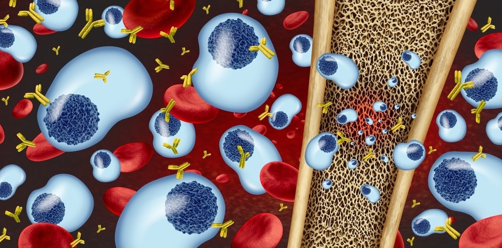
Image Credit: Lightspring/Shutterstock.com
Establishing diagnostic criteria
While routine blood tests can flag early indicators of MM, such as anemia or elevated total protein levels, further investigations are required to accurately detect the production of a monoclonal protein, such as serum and urine electrophoresis, and sFLC analysis. Additionally, advanced imaging techniques, such as MRI and PET scans, may be employed to identify bone lesions or other organ involvement.
The International Myeloma Working Group (IMWG) first established formal diagnostic criteria for multiple myeloma (MM) and its precursor condition in 2003,17 with an update in 2014 that introduced new diagnostic markers to support earlier and more accurate detection.5 In 2009, the IMWG also recommended replacing traditional urine analysis with serum free light chain (sFLC) testing in all myeloma investigations.18 This guidance has since been echoed by other bodies, including NICE (NG35)19 in 2016 and the British Society for Haematology (BSH)20 in 2022, both of which support sFLC testing as a preferred alternative to urine testing.
The diagnosis of MGUS is made when serum paraprotein levels are below 30 g/L, abnormal plasma cells make up less than 10 % of bone marrow cells, and there is no evidence of organ damage. In contrast, a diagnosis of active MM requires the presence of at least 10 % clonal plasma cells in the bone marrow along with one or more myeloma-defining events, as outlined in the CRAB or SLiM criteria.
The SLiM criteria identifies MM in patients who meet any of the following:
- ≥60 % clonal plasma cells in the bone marrow
- A serum involved/uninvolved free light chain (FLC) ratio of ≥100, where the involved FLC is at least 100 mg/L
- Imaging evidence of one or more focal lesions measuring 5 mm or larger
Together, these evolving diagnostic standards highlight the importance of integrating advanced testing methods to support early and accurate diagnosis.
Improving diagnosis with sFLC assays
Traditionally, serum and urine protein electrophoresis have been the mainstay methods for detecting paraproteins. However, urine electrophoresis has notable limitations - it lacks sensitivity, depends on the presence of free light chains in the urine, and is contingent on patient compliance in providing samples. These issues often lead to missed or delayed detection of light chain paraproteins, making the method unreliable for timely diagnosis.
To improve diagnostic accuracy, the IMWG (Dispenzieri 2009)18, the National Institute for Health and Care Excellence (NICE) 201619, and the BSH in 202220, updated their recommendations advising the replacement of urine electrophoresis with sFLC assays. These blood-based tests offer higher sensitivity and precision and, because blood samples are routinely collected from all patients suspected of MM, they eliminate logistical challenges associated with urine collection.
Unlike urine tests, sFLC assays can detect monoclonal free light chains in up to 97 percent of MM patients, making them especially useful in identifying light chain and non-secretory myeloma - forms that often evade traditional methods due to low biomarker levels or lack of secretion.21,22
Diagnosing non-secretory myeloma is particularly difficult because it features low levels of paraprotein in the serum, often below the detection capabilities of standard tests. sFLC assays overcome this by quantifying free κ and λ light chains in the blood and identifying abnormal κ/λ ratios - a strong indicator of clonal plasma cell activity. These abnormal ratios have been found in around 70 percent of non-secretory myeloma cases, making sFLC a critical diagnostic tool for detecting this elusive subtype even when traditional paraprotein tests fail.23
Despite these proven benefits, sFLC assays are still not widely used in the early detection of MM. Many UK hospitals remain confined to monitoring disease progression, despite NICE, the British Society for Haematology (BSH), and the International Myeloma Working Group (IMWG) advocating their use for initial diagnosis.18-20 This highlights a significant gap between clinical guidelines and real-world practices.
Frustratingly, budgetary constraints are often responsible for preventing labs from adopting sFLC assays for early diagnosis of MM, despite the best practice guidelines and clear evidence from trusts across the UK that have gone down this path. Healthcare systems and policymakers have the power to overcome this hurdle but need to recognize the long-term savings and key patient benefits associated with early diagnosis and realign budgets accordingly. Reallocating funds to prioritize prompt diagnosis is a complex but necessary step toward creating a more effective and patient-centric healthcare system.
Encouragingly, many UK labs are already equipped to drive this transformation. Freelite® assays - performed on the Optilite®* automated analyzer - are already widely used for monitoring MM patients, yet their diagnostic potential remains largely underutilized. These tests were introduced in 2001 as the first commercially available automated immunoassay of their kind, and they were purposely designed to enhance diagnostic precision and reduce delays.
Expanding the application of Freelite assays from monitoring to investigative purposes would therefore require minimal additional investment, as the necessary infrastructure is already in place. This small shift could yield major improvements, since automated testing enhances precision, reduces manual errors, streamlines workflows, and enables earlier detection, helping to minimise the costs and clinical burden of late-stage MM.
Spearheading the future of myeloma diagnostics
There has been significant progress in understanding and diagnosing MM, but healthcare systems have been slow to adopt best practices for early detection. Tools like sFLC assays, routinely used for monitoring, remain underutilized for diagnosis, despite their potential to significantly improve patient outcomes and reduce costs associated with late-stage disease. Achieving meaningful change requires healthcare systems to prioritize timely detection, improve cross-department collaboration, and shift budgets toward preventive care.
Industry leaders such as Binding Site, part of Thermo Fisher Scientific, are instrumental in this shift, providing both advanced diagnostics and the advocacy needed to support widespread adoption, ultimately delivering better patient care. Embracing early diagnosis as the optimal solution for managing MM enables stakeholders to create a future where patients receive timely, life-enhancing care, while also alleviating the burden of late-stage disease on already overstretched healthcare systems.
*Optilite and Freelite are registered trademarks of The Binding Site Group Ltd (Birmingham, UK) in certain countries.
Not for use in the United States or China.
References
- NationalInstituteforHealthandCareExcellence (2025). CKS is only available in the UK | NICE. (online) NICE. Available at: https://cks.nice.org.uk/topics/multiple-myeloma/background-information/prevalence (Accessed 18 Jul. 2025).
- NationalInstituteforHealthandCareExcellence (2025). CKS is only available in the UK | NICE. (online) NICE. Available at: https://cks.nice.org.uk/topics/multiple-myeloma/diagnosis/when-should-i-suspect-multiple-myeloma/https://cks.nice.org.uk/topics/multiple-myeloma/background-information/prevalence (Accessed 18 Jul. 2025).
- Cancer Research UK. Myeloma symptoms | Cancer Research UK. (online) Available at: https://www.cancerresearchuk.org/about-cancer/myeloma/symptoms.
- Myeloma UK. Ask the Nurse: Myeloma and blood cells. (online) Available at: https://www.myeloma.org.uk/library/ask-the-nurse-myeloma-and-blood-cells/.
- Rajkumar, S.V., et al. (2014). International Myeloma Working Group updated criteria for the diagnosis of multiple myeloma. The Lancet. Oncology, 15(12), pp.e538-548. https://doi.org/10.1016/S1470-2045(14)70442-5.
- Myeloma UK. MGUS. (online) Available at: https://www.myeloma.org.uk/understanding-myeloma/related-conditions/mgus/.
- Ji, M., et al. (2023). Mortality in the US Populations With Monoclonal Gammopathy of Undetermined Significance. JAMA Oncology, 9(9), pp.1293–1293. https://doi.org/10.1001/jamaoncol.2023.2278.
- Hevroni, G., et al. (2024). From MGUS to multiple myeloma: Unraveling the unknown of precursor states. Blood Reviews, pp.101242–101242. https://doi.org/10.1016/j.blre.2024.101242.
- International Myeloma Foundation (2021). Types of Myeloma. (online) International Myeloma Foundation. Available at: https://www.myeloma.org/types-of-myeloma.
- Pandey, S. and Kyle, R.A. (2013). Unusual myelomas: a review of IgD and IgE variants. Oncology (Williston Park, N.Y.), (online) 27(8), pp.798–803. Available at: https://pubmed.ncbi.nlm.nih.gov/24133829/.
- Agbuduwe, C., et al. (2022). Clinical characteristics and outcomes of IgD myeloma: experience across UK national trials. Blood Advances, 6(17), pp.5113–5123. https://doi.org/10.1182/bloodadvances.2022007608.
- Beatriz Nafría Jiménez and Raquel Oliveros Conejero (2021). IgE multiple myeloma: detection and follow-up. Advances in Laboratory Medicine / Avances en Medicina de Laboratorio, (online) 3(1), pp.79–84. https://doi.org/10.1515/almed-2021-0087.
- Brioli, A., et al. (2014). Serum free immunoglobulin light chain evaluation as a marker of impact from intraclonal heterogeneity on myeloma outcome. Blood, 123(22), pp.3414–3419. https://doi.org/10.1182/blood-2013-12-542662.
- Leung, N., et al. (2008). Improvement of cast nephropathy with plasma exchange depends on the diagnosis and on reduction of serum free light chains. Kidney International, (online) 73(11), pp.1282–1288. https://doi.org/10.1038/ki.2008.108.
- Cancer Research UK (2023). Survival | Myeloma | Cancer research UK. (online) Cancer Research UK. Available at: https://www.cancerresearchuk.org/about-cancer/myeloma/survival.
- Bong and Esa, E. (2023). Molecular genetic aberrations in the pathogenesis of multiple myeloma. Asian Biomedicine, (online) 17(4), pp.152–162. https://doi.org/10.2478/abm-2023-0056.
- Criteria for the classification of monoclonal gammopathies, multiple myeloma and related disorders: a report of the International Myeloma Working Group. (2003). British Journal of Haematology, (online) 121(5), pp.749–757. https://doi.org/10.1046/j.1365-2141.2003.04355.x.
- Dispenzieri, A., et al. (2008). International Myeloma Working Group guidelines for serum-free light chain analysis in multiple myeloma and related disorders. Leukemia, 23(2), pp.215–224. https://doi.org/10.1038/leu.2008.307.
- NICE (2016). Overview | Myeloma: diagnosis and management | Guidance | NICE. (online) NICE. Available at: https://www.nice.org.uk/guidance/ng35.
- Sive, J., et al. (2021). Guidelines on the diagnosis, investigation and initial treatment of myeloma: a British Society for Haematology/UK Myeloma Forum Guideline. British Journal of Haematology, 193(2), pp.245–268. https://doi.org/10.1111/bjh.17410.
- Katzmann, J.A., et al. (2009). Screening Panels for Detection of Monoclonal Gammopathies. Clinical Chemistry, 55(8), pp.1517–1522. https://doi.org/10.1373/clinchem.2009.126664.
- Dejoie, T., et al. (2016). Serum free light chains, not urine specimens, should be used to evaluate response in light-chain multiple myeloma. Blood, (online) 128(25), pp.2941–2948. https://doi.org/10.1182/blood-2016-07-726778.
- Drayson, M., et al. (2001). Serum free light-chain measurements for identifying and monitoring patients with nonsecretory multiple myeloma. Blood, 97(9), pp.2900–2902. https://doi.org/10.1182/blood.v97.9.2900.
Author biography
Dale Powner is the Country Manager for the UK, Ireland and Nordics at Binding Site, part of Thermo Fisher Scientific, a company that provides products and solutions for diagnosing and monitoring blood cancers and immune system disorders. Since joining the company in 2010, Dale has held diverse roles, from Medical Science Liaison to Business Development Manager. He now leads regional operations, focusing on advancing diagnostics, promoting early detection and improving patient outcomes, particularly in multiple myeloma and related conditions.
About Binding Site
Binding Site, Part of Thermo Fisher Scientific, is committed to improving patient lives worldwide through education, collaboration and innovation.
Binding Site provide diagnostic solutions that help doctors, clinicians and laboratory researchers across the globe diagnose and monitor blood cancers and immune system disorders.
Founded by researchers at the University of Birmingham, Binding Site has continued to build on its strong scientific foundations, supporting research and development within its field and responding to the changing needs of patients, researchers, and clinicians for over 30 years.
Sponsored Content Policy: News-Medical.net publishes articles and related content that may be derived from sources where we have existing commercial relationships, provided such content adds value to the core editorial ethos of News-Medical.Net which is to educate and inform site visitors interested in medical research, science, medical devices and treatments.