Cryopreservation is a widely utilized yet essential step in cell biology research, enabling the preservation of patient-derived samples, stem cells, immune cells, organoids, valuable cell lines, and other biological models destined for use in future experiments.
Once cells have been thawed, the initial question is often simply: “Are the cells alive?” This immediate post-thaw viability is a key quality check, with low viability potentially highlighting unsuitable freezing conditions, poor handling, or damage during storage and thawing.
Viability alone does not consistently answer the more important experimental question, however: “Are the thawed cells functionally recovered and suitable for reliable downstream use?”
Cryopreservation can impact cell attachment, proliferation, morphology, metabolism, differentiation capacity, marker expression, immune response, or assay performance, meaning that this distinction is especially important.
Successful cryopreservation must be defined as more than survival for complex or high-value cell models. Rather, this should mean recovery of the essential biological characteristics necessary for the next experiment.
Immediate post-thaw viability is an essential first checkpoint, but it does not always confirm experiment readiness. For sensitive or high-value cell models, cryopreservation success should be evaluated using recovery endpoints aligned with that match intended downstream application.
Viability’s history as the default metric
Post-thaw viability is commonly utilized because it is rapid, widely understood, and easy to quantify.
Methods such as automated cell counting, Trypan blue exclusion, and fluorescent live/dead stains provide researchers with an instant indication of apparent cell survival or membrane integrity.
These methods are limited, however, despite their usefulness. For example, most viability assays are not designed to measure whether cells have restored normal metabolism, whether immune cells are able to respond to stimulation, whether adherent cells can reattach efficiently, or whether stem cells have maintained the capacity to differentiate.
There is also a risk that these methods may not highlight delayed injury that only becomes apparent after several hours or days in culture.
A broad review of cell-specific considerations and cryopreservation principles highlights that both viability and functional assays are key to effectively evaluating post-thaw cell quality.1
Viability is, therefore, a required checkpoint, but not always a suitable endpoint.

Image Credit: NIPPON Genetics EUROPE GmbH
Differentiating between ‘alive’ and ‘experiment-ready’ cells
Immediate post-thaw viability is beneficial, but it cannot consistently predict whether cells will recover the required characteristics for downstream experiments.
Published studies demonstrate that cells may appear viable or recoverable via basic survival measures across different cell models.2,3,4,5,6,7 Later or model-specific readouts reveal changes in metabolism, immune subset composition, adhesion, differentiation potential, assay-relevant function, and 3D structure.
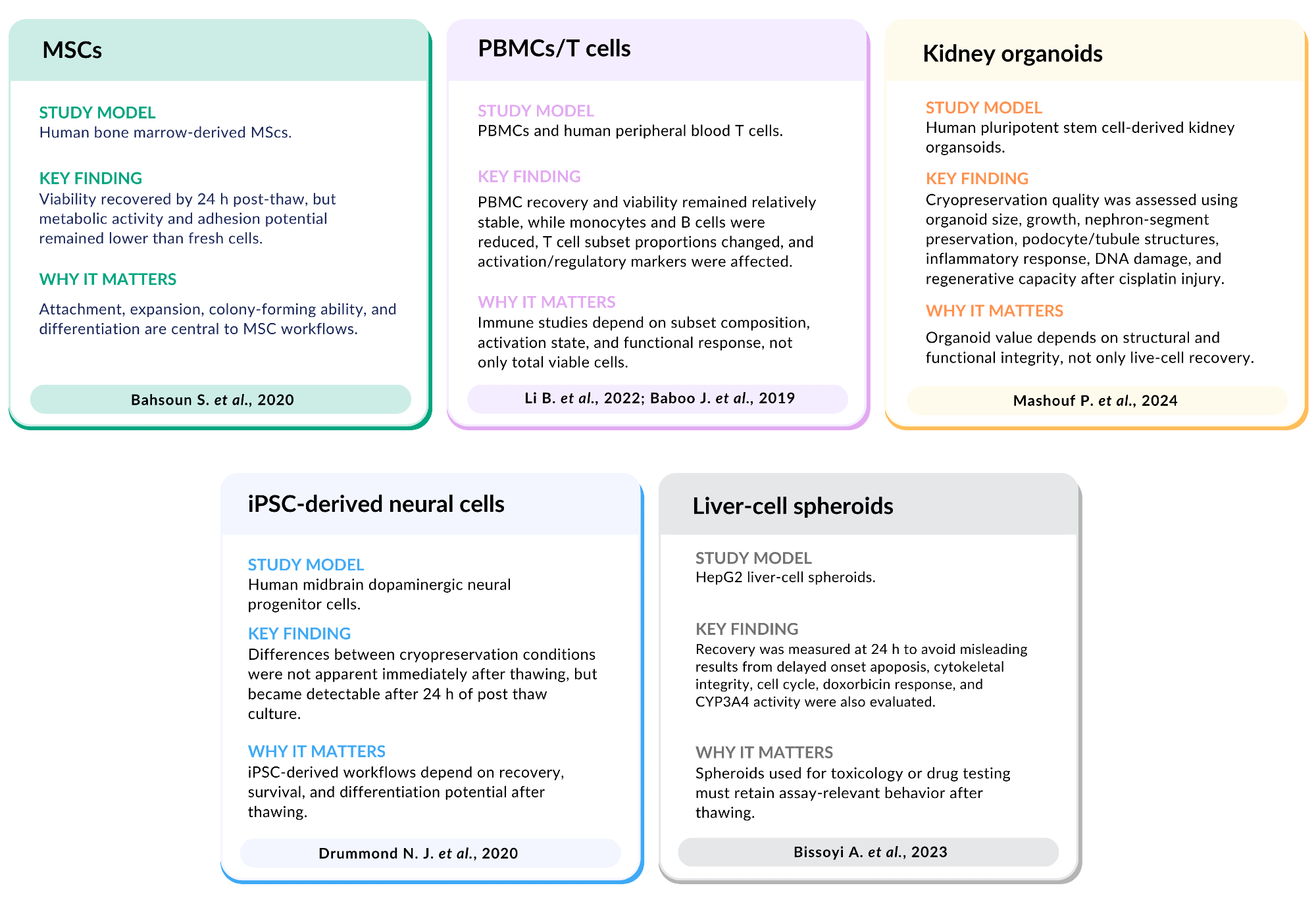
Image Credit: NIPPON Genetics EUROPE GmbH
These examples illustrate why ‘alive’ and ‘experiment-ready’ should be understood as different endpoints. Immediate viability verifies cells’ short-term survival after thawing, while functional recovery confirms that cells continue to exhibit the biological properties necessary for the intended assay, model, or workflow.
Functional recovery for different cell models
However, functional recovery should not be considered a single universal endpoint. The most relevant recovery readouts depend on the sensitivity of the cells, the biological model, and the downstream experiment in question.
For example, a viability assay can show that cells survived the freeze-thaw process, but this will not necessarily confirm that the recovered cells retain their relevant beneficial properties following freezing.
Functional recovery should, therefore, be defined in terms of the cells’ intended use.
For example, recovery may focus on attachment, morphology, and proliferation in routine adherent cultures,2 while this may depend on preserved subset composition and response to stimulation in immune cell workflows.3,4,12
Recovery may necessitate the retention of maturation state, phenotype, or differentiation potential in stem cell or iPSC-derived workflows,5,9 while this could depend on 3D architecture, population diversity, molecular profile, or assay performance in spheroids, organoids, and patient-derived samples.6,7,8,11
Source: NIPPON Genetics EUROPE GmbH
| Cell model |
Why viability alone may be incomplete |
Functional recovery may include |
| MSCs |
Cells may survive thawing but still show impaired attachment, spreading, metabolic activity, or expansion. For MSCs, this matters because attachment, colony-forming ability, and differentiation are
often central to downstream use. |
Attachment, morphology, spreading, proliferation, metabolic activity, colony-forming ability, differentiation potential |
PBMCs/T
cells |
Overall viability may not reflect changes in immune subset composition, activation state, apoptosis, proliferation, or response to stimulation. This is important when cryopreserved cells are used for immunophenotyping, cytokine assays, activation studies, or functional immune assays. |
Subset preservation, activation markers, apoptosis, proliferation after stimulation, cytokine production, suppressive function |
iPSC-derived
cells |
Cells may survive thawing but lose recovery capacity, phenotype, maturation status, or differentiation potential. For iPSC-derived models, the downstream value often depends on whether the cells can continue along the intended lineage or retain model-specific function. |
Recovery after 24 h, marker expression, differentiation potential, maturation markers, neurite outgrowth or model-specific morphology, functional assays |
Organoids/
spheroids |
Viable cells may remain after thawing, but 3D architecture, cell-cell organization, tissue-like morphology, growth behavior, or assay response may be compromised. This matters because 3D models are often used to approximate tissue biology more closely than 2D cultures. |
Structural recovery, morphology, regrowth, tissue-specific markers, cell organization, drug-response behavior, toxicology readouts |
Patient-derived
samples |
Viability alone may not show whether patient-specific biology remains usable for profiling, culture initiation, organoid generation, or functional testing. These samples are often limited and high value,
so biological usefulness after thawing is
the key endpoint. |
Culture initiation, molecular profiling, cell population preservation, histological features, genomic consistency, drug-response patterns |
A practical framework
Three sequential questions should be asked when evaluating the success of cryopreservation:
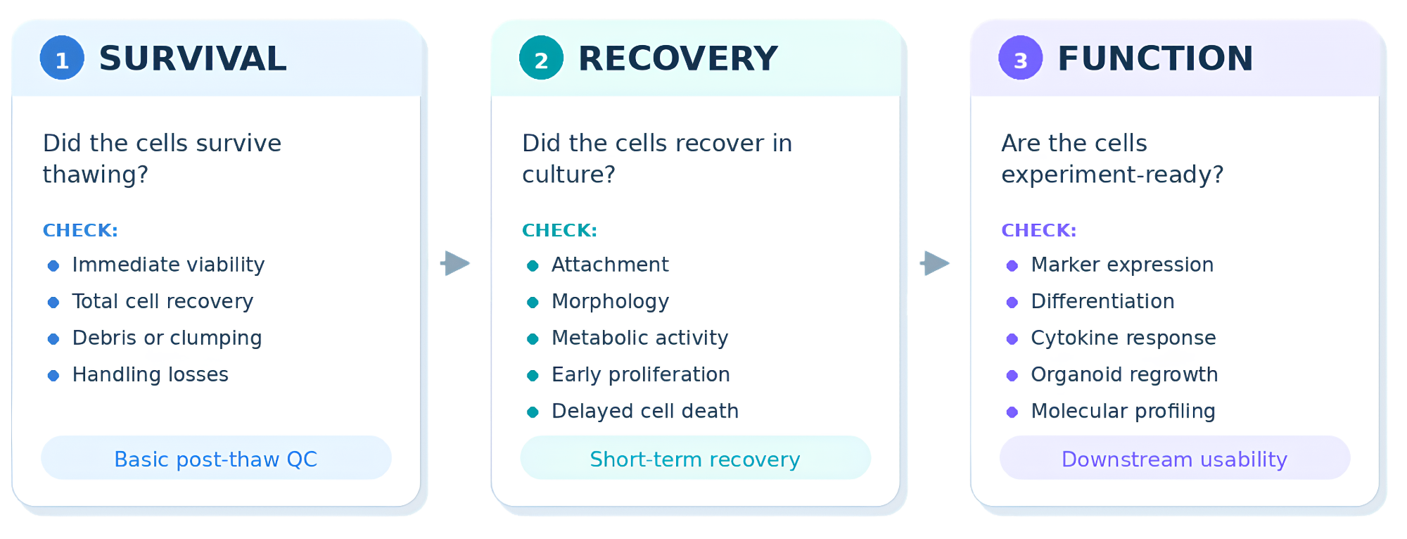
Image Credit: NIPPON Genetics EUROPE GmbH
This framework helps to distinguish three different levels of post-thaw quality. A sample showing acceptable immediate viability may pass the first checkpoint, but this would still require additional recovery or functional testing before being considered experiment-ready.
It is important to select the most relevant endpoints in line with the cell model and the planned downstream application.
Bambanker™ cryopreservation media
Bambanker™ is an FBS-free cryopreservation media that has been specifically developed to support efficient, reproducible cell freezing workflows.
Its streamlined protocol helps limited serum-containing preparation requirements, allowing laboratories to reduce hands-on handling time, simplify cryopreservation logistics, and minimize workflow-associated costs.
Cryopreservation outcomes are shaped by both the freezing medium and the workflow employed in the cells’ preparation, freezing, storage, and thawing.
Conventional or stepwise freezing protocols may require preparing serum-containing freezing mixtures, intermediate handling, controlled cooling steps, gradual addition of cryoprotectant, or equipment-dependent freezing procedures.
These workflows can be appropriate for regulated protocols and specific cell types, but they also increase both the number of variables that must be controlled and the required hands-on time.
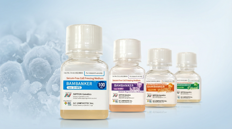
Image Credit: NIPPON Genetics EUROPE GmbH
Bambanker™ has been developed to simplify this process. Cells are suspended in Bambanker™ and frozen in standard workflows, helping to minimize handling complexity. Bambanker™ Direct allows cells to be frozen without washing or centrifugation, further simplifying the freezing process.
Source: NIPPON Genetics EUROPE GmbH
| Product |
Best for |
Key differentiator |
When to choose |
Bambanker™
Standard |
|
Versatile medium
Broad-use |
Default option for routine cell banking |
| Bambanker™ hRM |
- ES (Embryonic Stem Cells)
- iPS (induced Pluripotent Stem Cells)
|
Contains human
serum albumin |
Animal-component–free approach is preferred |
| Bambanker™ DMSO Free |
|
No DMSO |
DMSO is undesirable (e.g., protocols minimizing DMSO exposure) |
| Bambanker™ Direct |
- Hybridoma cells
- High-throughput (HTP) applications
|
No centrifugation of
cells is required |
Time-efficient freezing workflow, especially on high-throughput freezing days |
All Bambanker™ types are serum-free, ready-to-use cryopreservation media.
Functional recovery with Bambanker™
Bambanker™ has been utilized in published workflows where post-thaw performance was evaluated beyond immediate viability.
These important examples link cryopreservation success with the recovered cells’ biological purpose in terms of growth, marker expression, secretion activity, morphology, molecular profiling, or in vivo function.
Post-thaw outcomes are reliant on the entire freeze-thaw workflow, meaning that these examples should be interpreted as publication-supported use cases as opposed to universal performance claims for every cell type.
iPSC-derived dopaminergic neurospheres
Hiramatsu S. et al. used Bambanker™ hRM together with a Proton Freezer system in order to investigate the cryopreservation of human iPSC-derived dopaminergic neurospheres for clinical application.9
This study is a robust example of functional recovery-focused cryopreservation (Figure 1) because the authors assessed multiple downstream readouts, including dopamine secretion, dopaminergic marker expression, transplantation outcomes, and electrophysiological activity in 6-OHDA-lesioned rats.
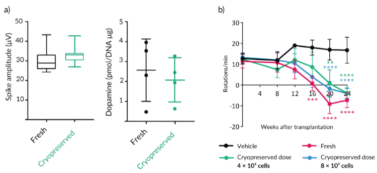
Figure 1. Functional recovery of iPSC-derived dopaminergic neurospheres after cryopreservation with Bambanker™. a) Cryopreserved iPSC-derived dopaminergic neurospheres showed electrophysiological activity and dopamine content in the same range as fresh neurospheres. Spike amplitude was comparable between fresh and cryopreserved spheres, and dopamine levels were maintained after cryopreservation. b) After transplantation into 6-OHDA-lesioned rats, fresh and cryopreserved dopaminergic neurospheres reduced abnormal rotational behavior over time compared with the vehicle control. Cryopreserved spheres showed a dose-dependent functional effect, with both tested cryopreserved cell doses contributing to reduced rotations after transplantation. **p < 0.01; ***p < 0.001; ****p < 0.000. Image Credit: Modified from Hiramatsu S. et al., 2022
The study shows that cryopreserved neurospheres demonstrated favorable post-thaw viability, as well as comparable dopamine secretion, dopaminergic marker expression, and electrophysiological activity to fresh spheres.
It was observed that the cryopreserved cells survived after transplantation, differentiating into mature dopaminergic neurons, and exhibited improved abnormal rotational behavior in the rat model.
These results highlight why solely relying on survival as an endpoint would not have been appropriate for this function-sensitive iPSC-derived model.
Bambanker™ has also been assessed in cultured human corneal endothelial cells. Okumura and their team screened an array of cryopreservation reagents, identifying Bambanker™ hRM as suitable for preserving clinical-grade human corneal endothelial cells.
Their study showed that cells cryopreserved with Bambanker™ hRM demonstrated post-thaw growth behavior and cell density comparable to non-preserved control cells following 28 days of culture (Figure 2a).
The cells also formed a monolayer sheet-like structure while retaining key corneal endothelial markers, including N-cadherin, ZO-1, and Na+/K+-ATPase (Figure 2b and Figure 2c).10
These results are applicable to functional recovery because it is important that preserved cells maintain growth, morphology, phenotype, and viability.
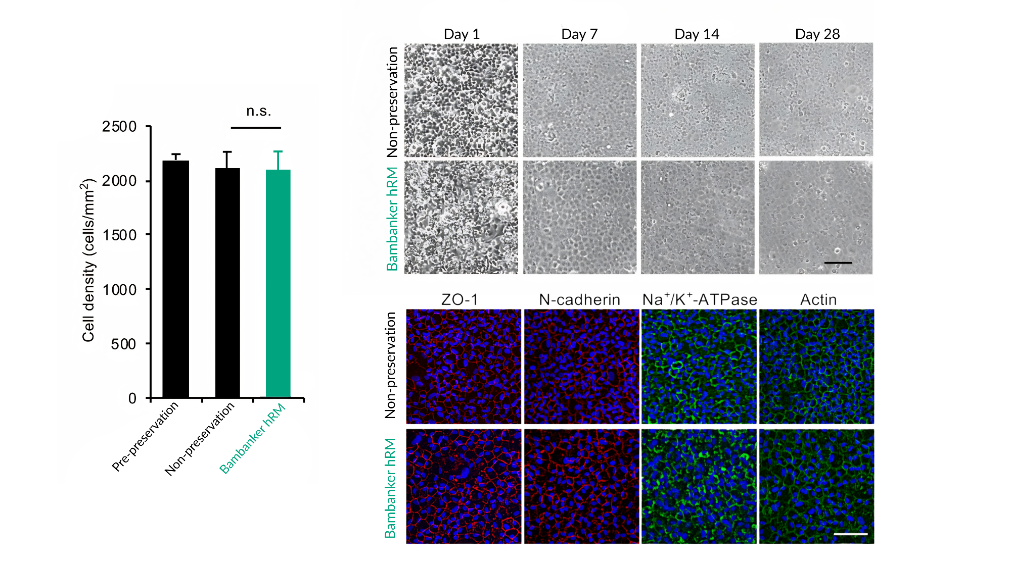
Figure 2. Post-thaw recovery of cultured human corneal endothelial cells cryopreserved with Bambanker™ hRM. a) Cell density after 28 days of culture was comparable between pre-preservation cells, non-preserved control cells, and cells cryopreserved with Bambanker™. The difference between non-preserved control cells and Bambanker™-preserved cells was not statistically significant. b) Phase-contrast images show that Bambanker™-preserved cells recovered over time in culture and formed a monolayer with morphology comparable to non-preserved control cells by day 28. c) Immunostaining showed that Bambanker™-preserved cells retained key human corneal endothelial cell markers, including ZO-1, N-cadherin, and Na+/K+- ATPase, with actin staining also shown. n.s. indicates no statistically significant difference. Image Credit: Modified from Okumura N. et al., 2019
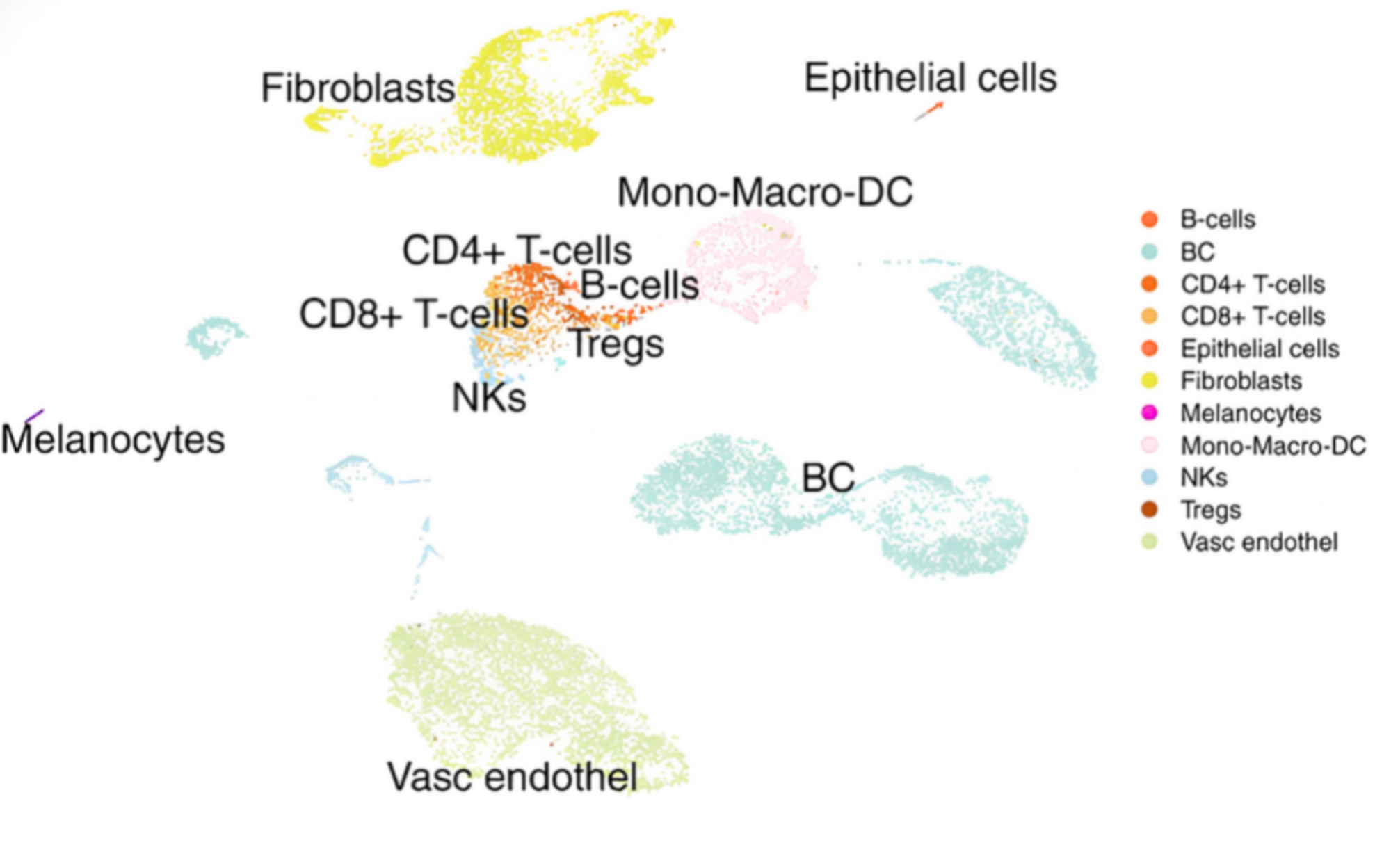
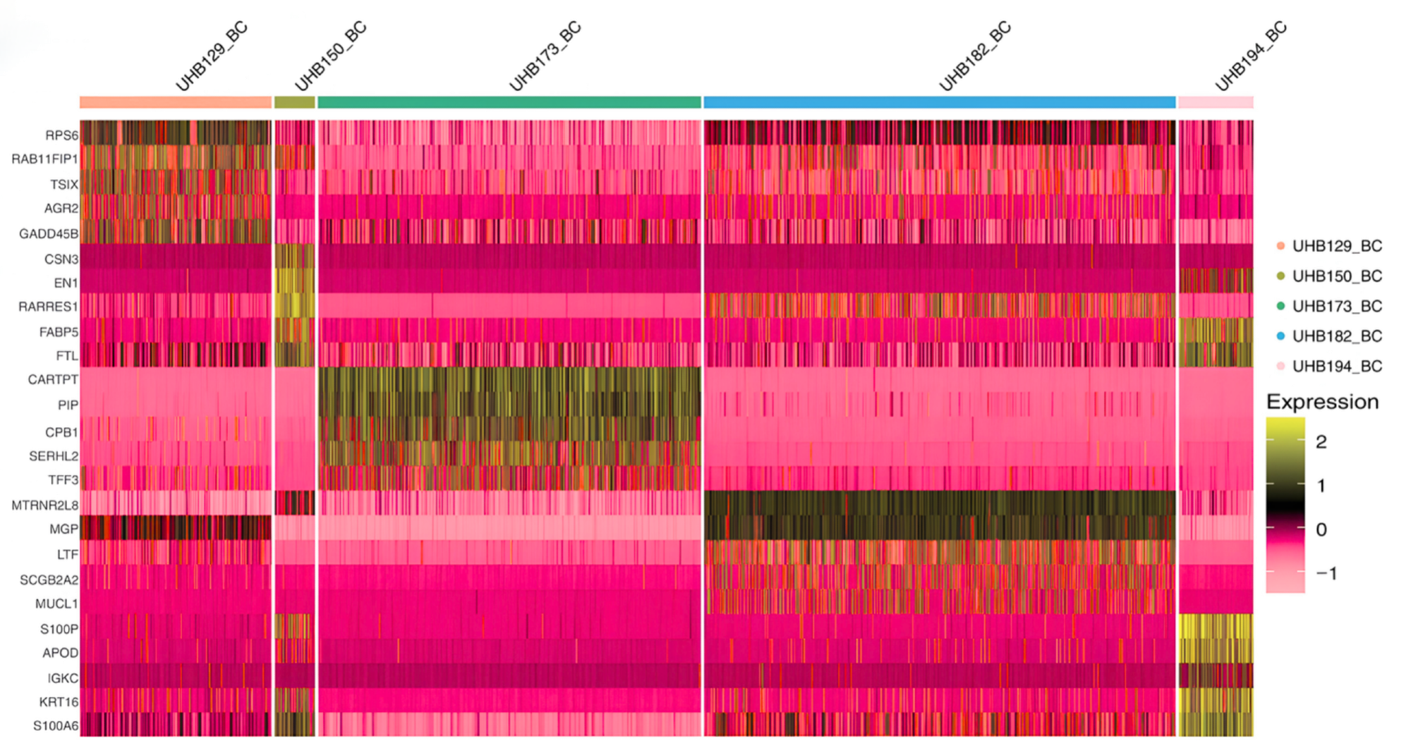
Figure 3. Single-cell profiling of patient-derived breast cancer tissue after slow-freezing in Bambanker™. a) Single-cell clustering identified multiple cell populations, including breast cancer cells, fibroblasts, immune cell subsets, epithelial cells, endothelial cells, melanocytes, and other stromal or immune populations. b) Marker-expression analysis showed breast cancer-relevant gene-expression patterns across recovered breast cancer cell populations. Together, these panels illustrate that Bambanker™-preserved patient-derived tissue could support downstream single-cell cellular profiling after thawing. Image Credit: Modified from Restivo G. et al., 2022
Another example of post-thaw success depending on preserving material for downstream analysis, as opposed to solely measuring viability, was provided by a study of patient-derived breast cancer tissue by Restivo G. et al.
The researchers utilized Bambanker™ in order to slow-freeze primary breast cancer tissue pieces prior to thawing, enzymatic digestion, and single-cell RNA sequencing.11
The recovered tissue supported single-cell clustering of multiple breast cancer-associated cell populations (Figure 3), including breast cancer cells, fibroblasts, epithelial cells, endothelial cells, immune cell subsets, and other stromal or immune populations.
Analysis of marker expression further highlighted the presence of breast cancer-relevant gene-expression patterns across recovered breast cancer cell populations. This study is relevant to functional recovery because the cryopreserved sample’s value was determined by whether it could still support biologically meaningful cellular profiling following thawing.
Conclusion
Immediate post-thaw viability continues to be a significant quality metric in cryopreservation, but it is important that this is not considered the only definition of success.
Published studies incorporating MSCs, PBMCs, T cells, and other systems demonstrate cryopreservation’s potential to impact key biological characteristics after the viability count is complete.2,3,4,12
The more useful question for contemporary cell culture workflows is whether cells are experiment-ready after thawing, particularly those workflows involving immune cells, stem cells, patient-derived samples, organoids, or function-sensitive assays.
Bambanker™ has been reported in peer-reviewed workflows across a diverse range of cell and sample types, supporting this broader conversation. Bambanker™-specific publications include numerous examples where post-thaw recovery was evaluated beyond simple survival.
The practical takeaway for researchers is clear: select and assess cryopreservation workflows based on the most relevant recovery endpoints for the next experiment.
References and further reading
- Whaley, D., et al. (2021). Cryopreservation: An Overview of Principles and Cell-Specific Considerations. Cell Transplantation, 30, p.096368972199961. DOI:10.1177/0963689721999617. https://journals.sagepub.com/doi/10.1177/0963689721999617.
- Bahsoun, S., Coopman, K. and Akam, E.C. (2020). Quantitative assessment of the impact of cryopreservation on human bone marrow-derived mesenchymal stem cells: up to 24 h post-thaw and beyond. Stem Cell Research & Therapy, 11(1). DOI:10.1186/s13287-020-02054-2. https://link.springer.com/article/10.1186/s13287-020-02054-2.
- Li, B., et al. (2022). Comprehensive evaluation of the effects of long-term cryopreservation on peripheral blood mononuclear cells using flow cytometry. BMC Immunology, 23(1). DOI:10.1186/s12865-022-00505-4. https://link.springer.com/article/10.1186/s12865-022-00505-4.
- Baboo, J., et al. (2019). The Impact of Varying Cooling and Thawing Rates on the Quality of Cryopreserved Human Peripheral Blood T Cells. Scientific reports, 9(1), p.3417. DOI:10.1038/s41598-019-39957-x. https://www.nature.com/articles/s41598-019-39957-x.
- Drummond, N.J., et al. (2020). Cryopreservation of Human Midbrain Dopaminergic Neural Progenitor Cells Poised for Neuronal Differentiation. Frontiers in Cell and Developmental Biology, 8. DOI:10.3389/fcell.2020.578907. https://www.frontiersin.org/journals/cell-and-developmental-biology/articles/10.3389/fcell.2020.578907/full.
- Parham Mashouf, et al. (2024). Cryopreservation of human kidney organoids. Cellular and Molecular Life Sciences, 81(1). DOI:10.1007/s00018-024-05352-7. https://link.springer.com/article/10.1007/s00018-024-05352-7.
- Bissoyi, A., et al. (2023). Cryopreservation of Liver-Cell Spheroids with Macromolecular Cryoprotectants. ACS Applied Materials & Interfaces. DOI:10.1021/acsami.2c18288. https://pubs.acs.org/aamick/article/15/2/2630/1243689/Cryopreservation-of-Liver-Cell-Spheroids-with.
- Chen, P., et al. (2025). Establishing a cryopreserved biobank of living tumor tissues for drug sensitivity testing. Bioactive Materials, 46, pp.582–596. DOI:10.1016/j.bioactmat.2024.09.008. https://www.sciencedirect.com/science/article/pii/S2452199X24003955?via%3Dihub.
- Hiramatsu, S., et al. (2022). Cryopreservation of Induced Pluripotent Stem Cell-Derived Dopaminergic Neurospheres for Clinical Application. Journal of Parkinson’s Disease, pp.1–14. DOI:10.3233/jpd-212934. https://journals.sagepub.com/doi/10.3233/JPD-212934.
- Okumura, N., et al. (2019). Feasibility of a cryopreservation of cultured human corneal endothelial cells. PLOS ONE, 14(6), p.e0218431. DOI:10.1371/journal.pone.0218431. https://journals.plos.org/plosone/article?id=10.1371/journal.pone.0218431.
- Restivo, G., et al. (2022). Live slow-frozen human tumor tissues viable for 2D, 3D, ex vivo cultures and single-cell RNAseq. Communications Biology, 5(1). DOI:10.1038/s42003-022-04025-0. https://www.nature.com/articles/s42003-022-04025-0.
- Jantet-Blaudez, F., et al. (2025). Evaluation of the viability and functionality of human peripheral blood mononuclear cells cryopreserved up to 2 years in animal-protein-free freezing media compared to the FBS-supplemented reference medium. Frontiers in Immunology, 16. DOI:10.3389/fimmu.2025.1627973. https://www.frontiersin.org/journals/immunology/articles/10.3389/fimmu.2025.1627973/full.
Acknowledgments
Produced from materials originally authored by NIPPON Genetics EUROPE GmbH.
About NIPPON Genetics EUROPE GmbH
Our contribution to your success – we are committed to ensuring that you can achieve your goals!
NIPPON Genetics EUROPE is a global biotech company founded in 2005 with its own research, development, and production based in Germany. By providing an extensive range of selected and groundbreaking products for the life science sector, we would like to be a reliable partner and contribute to achieving our customers' individual goals. The majority of our employees have a scientific background because we want to understand which problem solutions our customers care about.
Sponsored Content Policy: News-Medical.net publishes articles and related content that may be derived from sources where we have existing commercial relationships, provided such content adds value to the core editorial ethos of News-Medical.net, which is to educate and inform site visitors interested in medical research, science, medical devices, and treatments.