Digital radiography (DR) is increasingly used in radiology practice, as the trend towards paperless and filmless radiography advances. DR is based on the use of X-rays for data acquisition with direct or indirect conversion to electrical charge using various detection-charge conversion systems.
The advantages of digital radiography are many.

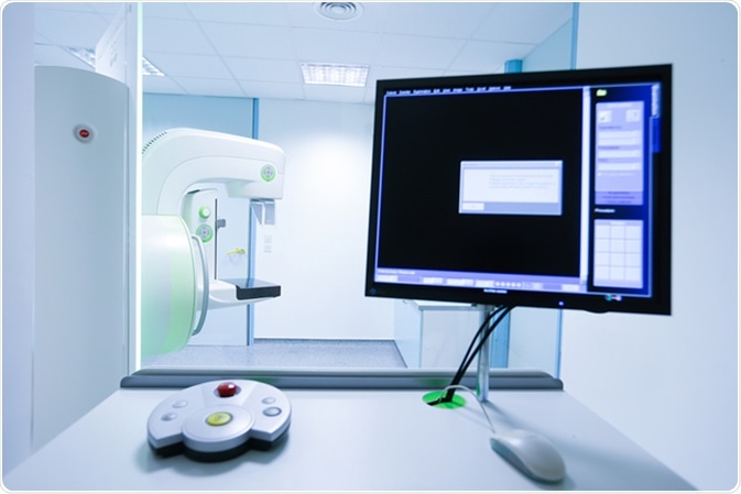
Mammography breast screening device in hospital. Image Credit: zlikovec / Shutterstock
Remote Secure Viewing
The outstanding benefit of adopting DR is the fully digitized picture archiving and communication system (PACS), which allows images to be archived on digital storage media and viewed by any physician in the network as necessary, at any time. This means that images obtained at a healthcare institution such as a hospital can be electronically distributed using the internet, and images cannot be lost accidentally.
Manual Image Acquisition
DR is suitable for image generation without automatic control of exposure, such as in bedside imaging. This is now possible because of developments that have brought down costs and increased availability of more versatile imaging units.
High Image Quality
Another DR advantage is its use of detectors that have markedly higher imaging power compared with conventional film or computerized imaging. Also, the separation of various steps needed to create the final image (image generation, processing, and documentation) is an advancement.
Of course, for maximal diagnostic utility, images resulting from DR must be optimized at each step in the process. If performance of any component is suboptimal, image quality will suffer.
Newer DR platforms provide better spatial resolution for anatomical features that are in close proximity to one another.
More Efficient Devices
X-ray detector efficiency can be measured using the detective quantum efficiency, or DQE, which denotes the level of efficiency with which photons are detected and the amount of noise in the signal following detection. The ideal DQE would be 100%, meaning that all X-ray photons were recorded in the image, and the noise was zero. However, DQEs are typically affected by incomplete X-ray absorption and internal noise originating from the device. DR systems have DQEs around 65%, which allow lower radiation dosages to be used (compared with traditional X-rays) with the same amount of quantum noise.
Reduced dose exposure occurs because of much lower failed exposures (which require repeated scans), and lower radiation dosages required to obtain individual images.
Speedy Imaging
Increased speed of imaging readouts is a great advantage that results from the integration of image acquisition, processing, and presentation in the same device.
DR is also capable of easier and more objective data collection from X-ray images, which allows for better interpretation and more accurate diagnosis.
Higher Efficiency of Workflow
Flat plane detectors also make it possible to view images within about 10 seconds. This high speed image presentation makes up for the high cost of installing the new technology in dedicated imaging facilities or hospitals that have heavy X-ray workloads. Patient throughput is significantly increased. Of course, necessary changes must be made to the workflow and the system of patient management to reap the economic benefits of the new technology.
Flat panel detectors are much better at imaging anatomic details clearly compared to conventional radiography. They also offer the greatest chance of lowering radiation exposure in any clinical setting.
Versatility
Several types of digital radiography detectors are available. Storage phosphor plates are compatible with all the current radiography units. Thus their use entails the least additional investment.
Flat plane detectors possess very good imaging properties even with low doses of radiation. This is because of the relatively thick scintillator layer that allows the light produced by the phosphor to orient itself for travel towards the underlying layer of photodiodes at the point of production. Both cesium and gadolinium salts are used, but the latter is inexpensive and durable, being preferred for use in ward X-rays using mobile radiography equipment.
Higher Dynamic Range
Other advantages of DR include a higher dynamic range coupled with the possibility of reducing X-ray exposure at the level of the individual patient. This is especially true because these platforms have much better quantum efficiency. The dynamic range is the range of X-ray exposure which allows acquisition of an acceptable image capable of interpretation. A higher dynamic range allows different tissues to be differentiated on the same image without additional imaging, by means of the specific tissue absorptions. However, care must be taken to avoid over-exposure. With digital radiography, image quality is only improved by overexposure.
Further Reading
Last Updated: Nov 18, 2018