Introduction to Gastroschisis
Etiology and pathophysiology
Gastroschisis diagnosis
Management approaches and prognosis
References
Further reading
Introduction to Gastroschisis
Gastroschisis (Greek word for “abdominal cleft”) refers to a usually small (<4cm) full-thickness paraumbilical (usually right-sided) congenital disability of the abdominal wall, allowing abdominal contents uncovered by any peritoneal membrane or sac to protrude through a gap in the abdominal wall (herniation of the fetal midgut). The herniated contents get exposed to the body's exterior and are susceptible to infection. Infants with gastroschisis are more likely to develop intrauterine growth restrictions and have preterm births.
In gastroschisis, herniation of other organs, such as the stomach, liver, spleen, or genitourinary tract, may occur but is uncommon. Long-term complications of gastroschisis include bowel dysmotility, short gut syndrome, and complications from long-term total parenteral nutrition, including liver failure.
The defect has been increasingly prevalent worldwide (especially in Japan, Australia, North, Central, and South America, and North-Central Europe) and usually does not present with concomitant chromosomal abnormalities. Most cases of gastroschisis have been reported in pregnancies of women under 20 years.
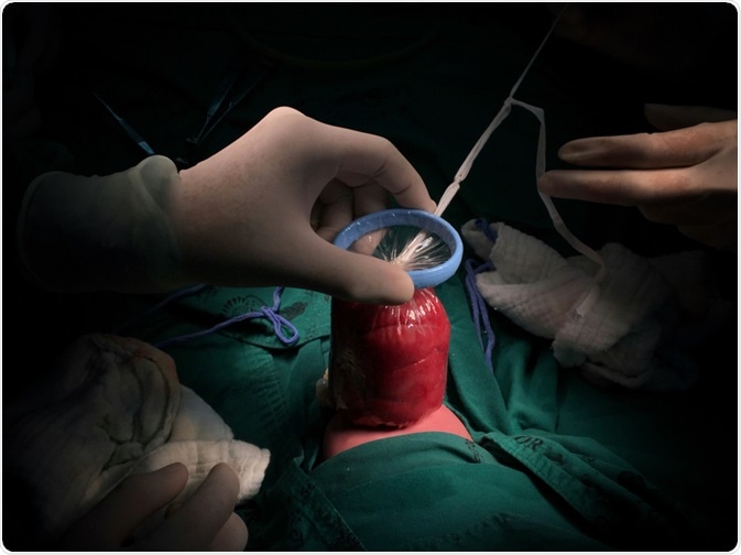
Image Credit: Paradee Siriboon / Shutterstock.com
Etiology and pathophysiology
The etiology of gastroschisis includes environmental causes, such as infection, nutrition, and medication use. Researchers have linked smoking, alcohol use, cocaine use, and the use of common medications such as acetaminophen, aspirin, ibuprofen, and pseudoephedrine. Researchers have also found a significant association between mothers who reported both urinary tract infection (UTI) and sexually transmitted infection (STI) in the month before conception or during the first trimester and fetal gastroschisis.
Further, there are isolated reports of gastroschisis associated with trisomies 13, 18, and 21 and monosomy 22. Gastroschisis has also been associated with gastrointestinal and central nervous system defects. About 10% of babies who have gastroschisis have intestinal stenosis or atresia that results from vascular insufficiency to the bowel at the time of gastroschisis development or, more commonly, from later volvulus or compression of the mesenteric vascular pedicle by a narrowing abdominal wall ring.
The abdominal wall is usually formed by infolding the cranial, caudal, and two lateral embryonic folds. As the abdominal wall forms, the rapid growth of the intestinal tract leads to its migration outside the abdominal cavity through the umbilical ring and into the umbilical cord during week 6 of gestation. By weeks 10 and 12, the abdominal wall is well formed, and the intestine returns to the abdominal cavity in a stereotypical pattern that results in normal intestinal rotation and subsequent fixation.
Gastroschisis has been regarded as a disruption (i.e., an abnormality produced after initial normal development) rather than a malformation (i.e., an abnormality occurring during early embryonic development). However, recent evidence indicates that gastroschisis may be a malformation. Several hypotheses have been proposed to describe events leading to the development of gastroschisis, all of which involve defective formation or disruption of the body wall.
Gastroschisis may be the consequence of the failure of one or more of the folds responsible for abdominal wall closure. This failure is the proposed mechanism for other ventral wall defects (e.g., ectopia cordis and cloacal exstrophy). The proposed mechanism states that the body fold failure impedes the merging of the yolk sac with the body stalk. As the gut development continues, part of the primary intestinal loop attached to the vitelline duct herniates through the body fold defect and into the amniotic cavity instead of the umbilical cord.
An alternative hypothesis has been proposed in which the primary intestinal loop herniates normally into the umbilical cord, with another part of the gut herniating through the unclosed portion of the ventral wall. Another hypothesis states that the determining defect in gastroschisis is the failure of the yolk sac and related vitelline structures to be incorporated into the umbilical stalk, which leads to the persistence of the vitelline duct and yolk sac outside the main body stalk and abdominal wall, whereas the lateral abdominal walls close normally.
The developing midgut has two points of egress from the abdominal cavity; this leads to abnormal herniation of the expanding midgut into the amniotic cavity and subsequent development of gastroschisis. Gastroschisis has been thought to result from an ischemic insult to the developing body wall. However, an alternative hypothesis that may account for some cases of gastroschisis is that the defect results from an early rupture of a hernia of the umbilical cord.
Events leading to the development of gastroschisis include (i) failure of embryonic mesenchyme to form the abdominal wall because of teratogen exposure, (ii) rupture of amnion around the umbilical ring during the time of physiologic herniation or later, (iii) abnormal involution of the right umbilical vein leading to weakening of the body wall and subsequent gut herniation and (iv) disruption of the right omphalomesenteric (vitelline or yolk sac) artery leading to infarction and necrosis at the base of the cord with subsequent body wall damage.
Other events include abnormal folding of the body wall resulting in a ventral body wall malformation leading to herniation of the fetal gut and the failure of the yolk sac and related vitelline structures to become incorporated into the body stalk, orphaning the vitelline duct and yolk sac outside both the main body stalk and the abdominal wall.
Gastroschisis diagnosis
Differential diagnoses of gastroschisis include omphalocele, ruptured omphalocele, limb–body wall complex, bladder exstrophy, cloacal exstrophy, ectopia cordis, pentalogy of Cantrell, umbilical cord cysts, and urachal abnormalities.
Generally, gastroschisis is detected by prenatal ultrasound (USG) screening beginning at the end of the first trimester and elevated maternal serum alpha-fetoprotein levels. The tests allow for antepartum diagnosis of gastroschisis with high accuracy and allow time to plan the birth and discuss the best way to manage the condition.
The classic USG appearance of fetal gastroschisis is free-floating loops of the bowel within the amniotic cavity. The exteriorized mass lacks a peritoneal covering and has a "cauliflower" appearance because the amniotic fluid between the loops of the bowel results in acoustic interfaces at both near and far bowel walls. Visualization of free-floating bowel loops is enhanced secondary to echogenic bowel wall edema, inflammation, and the dilated intestinal lumen. In addition, the stomach and intestine may dilate because of obstruction (volvulus, atresia or stenosis, or malrotation) or hypoperistalsis.
Management approaches and prognosis
There are presently no prenatal treatment options for fetal gastroschisis. Secondary to the exposure of the eviscerated bowel, the neonate is at a high risk of insensible losses of fluid and heat. The ABCs of resuscitation (Airway, Breathing, and Circulation) and serum glucose levels need to be maintained. Babies with gastroschisis have high fluid losses from evaporation and third-space losses and may require twice the maintenance volumes of fluids to maintain an adequate intravascular volume.
Gastroschisis requires prompt surgical management as soon as the baby is born. The type and extent of the operation depend on the number of intestines that reside outside the abdomen and the space available to fit the intestine back inside the abdominal cavity. The baby is usually anesthetized within 24 hours of birth, and an operation is performed to place the exposed intestines back inside the cavity and seal the hole in the abdominal wall.
Immediately after delivery, the herniated bowel is wrapped with sterile saline dressings and covered in plastic wrap. If pediatric surgery is not immediately available for evaluation, the neonate is placed on the right side to avoid kinking of the blood supply as it traverses through the abdominal defect. A nasogastric tube is placed to decompress the bowel maximally, and intravenous access is obtained for fluid resuscitation.
The preferable closure method for routine gastroschisis cases is by bedside placement of a Silastic spring-loaded silo. After the placement of the spring-loaded silo, the baby is evaluated further and cared for in the intensive care unit (ICU). With spontaneous diuresis, gastrointestinal tract decompression from above and below, and resolution of bowel wall edema, the volume of the exposed bowel in the bag markedly decreases in a short period. Among the advantages of this method are minimal fluid resuscitation requirements and no need for anesthesia or intubation.
Defects are usually reduced in one to three days. In some cases, placing the entire intestine back inside the cavity in one operation is impossible, and the procedure must be performed in stages.
The surgeon places a mesh over the intestine for a staged repair to contain and protect it. This is then suspended above the child, and gravity eventually moves the intestine back inside the abdomen. The mesh sack is tightened regularly until all of the intestines have moved back into the abdomen.
Gastroschisis can also be reduced via primary closure or staged closure. Once the reduction is performed, the skin defect may be closed with traditional suture closure or with a biologic closure using the remnant of the umbilical cord.
Overall, children born with this condition have an excellent prognosis. Today, the survival rate is around 90% to 95%, and most of the deaths are in patients with catastrophic bowel loss, sepsis, and the long-term complications of short-bowel syndrome.
Fetal Gastroschisis | FAQ's with Dr. Eric Jelin
References
Further Reading
Last Updated: Feb 29, 2024