This article is based on a poster originally authored by Anne J Hout, Nikki Claassen, Ilse H Wolters, Johanna FB Pagano, and Karthikeyan Devaraju.
Stable expression in gene therapies with transposons, lentiviral (LVV), and retroviral (RVV) vectors is achieved by integrating a therapeutic gene into the host's genome. LVV or RVV are predominantly used in cell and gene therapies, particularly in CAR/TCR-T (chimeric antigen receptor/T-Cell receptor) therapies.
These vectors typically follow randomized integration patterns, meaning genetic safety testing is essential to allay any concerns related to vector genotoxicity via activation of proto-oncogenes.
Vector integration sites (VIS) in the host genome must be identified precisely to validate the safe insertion of the therapeutic gene while avoiding therapy-induced oncogenesis
Materials and methods
- Guide RNAs (gRNA) were developed based on the conserved regions in the lentiviral vectors, enabling selective enrichment of regions of interest in background human DNA.
- Synthetic lentiviral construct and single viral integration cell lines were used alongside World Health Organization (WHO) reference materials at 10 integration sites.
- The MinION flow cells and relevant sequencing library preparation kit (V14 kit) for the MinION Mk 1D were acquired directly from Oxford Nanopore Technologies (OPT), UK.
- Standard pipelines were used for aligning sequences and enrichment. A custom script was developed for annotating insertion sites.
Results
- gRNAS were developed to target the vector sequences to enrich integration sites using CRISPR-Cas9. Oxford Nanopore’s Mk1D sequencer was used for subsequent sequencing.
- A synthetic DNA construct was used to verify the pipeline of the gRNA target sequences. All quality control criteria were met, and no errors occurred across sequencing runs. The base calling and resulting data were evaluated using a custom bioinformatics pipeline, which facilitated vector identification as displayed in Figure 6.
- The pipeline was then tested to validate VIS in WHO control materials as shown in Figure 7. The resulting data was assessed using the bioinformatics pipeline, which was then mapped to read the viral vector and human reference genomes. This determines the alignment breakpoints of the chimeric reads, which subsequently identifies integration sites.
- Using a combination of gRNAs in cis and trans, all 10 integrated sites of the WHO control sample were successfully enriched and detected. Subsequently, the pipeline was verified using a cell line with one lentiviral integration event (data not shown).
- This pipeline facilitates detection of integrated LVVs used in gene therapies (CAR/TCR-T and other gene therapies) at both the integration sites and vector sequences. The sensitivity range of the assay is still being evaluated.

Figure 1. VIS Assay Workflow for CRISPR-Cas9 targeted ONT sequencing. Image Credit: Cerba Research
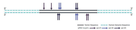
Figure 2. gRNA design on target sequences. Image Credit: Cerba Research
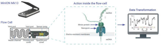
Figure 3. Snapshot of sequencing process inside a MinION flow cell and transformation of electrical signals to nucleotides. Image Credit: Cerba Research
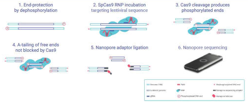
Figure 4. Assay workflow overview. The genomic DNA is processed and incubated with Ribo-Nucleo- Protein (RNP) complex to enrich insertion sites for nanopore sequencing. Image Credit: Cerba Research
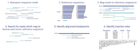
Figure 5. Data Analysis workflow for mapping the nanopore sequence reads to the vector and human genome sequences. The integration sites are identified by custom pipeline. Image Credit: Cerba Research
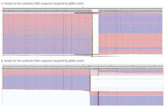
Figure 6. The above images show the reads from the synthetic DNA sequence screened for gRNAs targeted nanopore sequencing. A., shows data from gRNA set#1. B., shows the data gRNA set#3. The gRNA set#1 was better than gRNA set#3 with more reads (coverage). Image Credit: Cerba Research
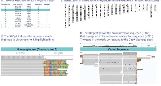
Figure 7. The above images and tables show the VIS data obtained with targeted nanopore sequencing. A., Lists the identified integration sites from the sequencing runs (n=4). The sites in blue box within the table shows the sites identified by many laboratories but not defined by W.H.O. B., visualizes the summary VIS data in depicting the integration sites within the chromosomes. C., shows the human genome sequence from the site highlighted in table A. D., shows the vector sequence identified from multiple genomic locations. Image Credit: Cerba Research
Conclusion
WHO controls were used to document and define all integration sites in this assay, with additional sites also identified as reported by other lab-based studies using the same WHO reference guide. It was determined that the pipeline can detect single integration events, which underscores the need to explore approaches to improve coverage.
Furthermore, future recommendations to advance the study include actively establishing the pipeline's sensitivity and seeking collaborations to test lentiviral vector therapies.
References and further reading
- Gilpatrick, T., et al. (2020). Targeted nanopore sequencing with Cas9-guided adapter ligation. Nature Biotechnology, 38(4), pp.433–438. DOI: 10.1038/s41587-020-0407-5. https://www.nature.com/articles/s41587-020-0407-5.
About Cerba Research

Cerba Research is a leading specialty laboratory services provider with the capacity and breadth of a global central laboratory network. Their highly qualified scientists provide insight on the latest biomarkers, assays and testing approaches and develop innovative solutions for unique challenges across all research phases, to pharmaceutical, biotechnology, medical device, government, public health, and CRO organizations.
Cerba Research’s extensive capability in laboratory testing and global logistics including Bioanalysis, Flow Cytometry, Histopathology, and Next-Generation Sequencing, enables them to drive operational agility at scale in a wide range of therapeutic areas, with recognized expertise in Virology, Immunology, Oncology and Cell & Gene Therapy.
Cerba Research is part of the Cerba HealthCare Group with 15,000 employees on five continents, driven to advance diagnosis and health.
For more information about Cerba Research, please visit cerbaresearch.com.
Sponsored Content Policy: News-Medical.net publishes articles and related content that may be derived from sources where we have existing commercial relationships, provided such content adds value to the core editorial ethos of News-Medical.net, which is to educate and inform site visitors interested in medical research, science, medical devices and treatments.
Last Updated: Jul 16, 2026
 Viroclinics-DDL changes name to Cerba Research