This article and associated images are based on a poster originally authored by Teresa Sorbo, Manuel Paina, Manuel Arcangeletti, Katja Blasi, Sara Martin, Donatella Bardelli, Paola Picardi, Fernanda Ricci, Silvia Cainarca, Jean-Francois Rolland, and Anna Mondini, and presented at ELRIG Drug Discovery 2025 in affiliation with Axxam S.p.A.
This poster is being hosted on this website in its raw form, without modifications. It has not undergone peer review but has been reviewed to meet AZoNetwork's editorial quality standards. The information contained is for informational purposes only and should not be considered validated by independent peer assessment.

Abstract
Lysosomal abnormalities are a hallmark of neurodegenerative diseases, with common features including altered lysosomal pH, impaired enzyme activity, and defects in lysosomal trafficking. TMEM175, a member of the transmembrane protein family, has garnered considerable attention for its critical role in lysosomal function.
This study investigates TMEM175 using a diverse set of methodologies, from high-throughput screening to detailed single-channel recordings on lysosomal membranes.
The team first generated stable cell lines expressing TMEM175 and identified potential modulators via a thallium fluorescence assay. Electrophysiological characterization followed, initially focusing on the plasma membrane and later extending to the lysosomal membrane.
Both manual and automated patch-clamp techniques were employed to precisely measure TMEM175-mediated ionic currents within isolated lysosomes. While manual patch-clamping provides valuable insights into organellar function, its labor-intensive nature and limited scalability for large-scale drug screening present significant challenges.
To address these, the team developed a robust lysosomal high-throughput screening (HTS) platform using the SyncroPatch system (Nanion), which dramatically enhances throughput by enabling compound testing on a 384-well array.
Lysosomal function was also evaluated in iPSC-derived dopaminergic neurons, providing a physiologically relevant model to study TMEM175 in the context of neurodegenerative diseases. This approach bridges mechanistic insights into TMEM175 function with disease-relevant cellular models, advancing understanding of its role in lysosomal health and its potential as a therapeutic target.
In summary, this comprehensive study highlights the significance of TMEM175 across various experimental paradigms, ranging from high-throughput platforms to intricate lysosomal analyses, thereby advancing translational research and enhancing our understanding of neurodegenerative disease mechanisms.
Summary
- Intracellular ion channels are crucial for various signaling pathways that govern both health and disease.
- Targeting lysosomal dysfunction has become a promising therapeutic approach for neurodegenerative disorders such as Parkinson's Disease (PD) and Amyotrophic Lateral Sclerosis (ALS), with a particular focus on understanding and modulating the function of lysosomal ion channels.
- The growing interest in intracellular ion channels as therapeutic targets underscores the need for high-throughput electrophysiology assays in the drug discovery process. However, the ability to gather data from native lysosomes has remained a significant challenge.
- This study addresses this gap by presenting data obtained from isolated lysosomes using an HTS automated patch-clamp device. An innovative lysosomal patch-clamp technique is applied to iPSC-derived neurons, providing new insights into lysosomal ion channel function and opening avenues for more efficient drug discovery.
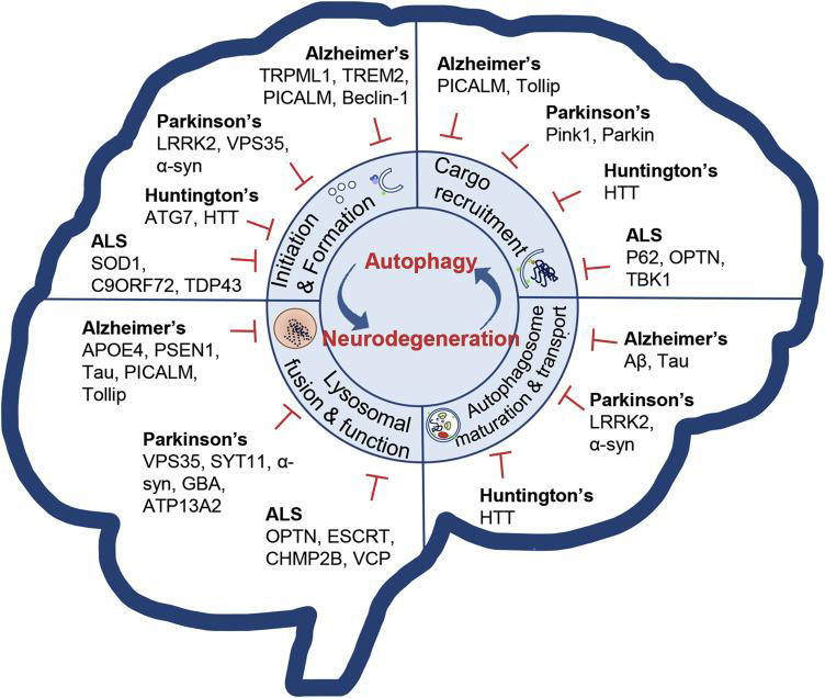
Image Credit: From: Fleming A. Neuron. 2022 Mar 16;110(6):935-966
This study is part of the AICoS project (CUP B79J23000340005) supported by the Italian Ministry of Economic Development (MISE) – Innovation Agreements D.M. 31/12/2021
LysoPatch – Automated mode
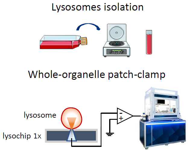
Image Credit: Image courtesy of Teresa Sorbo et al., in partnership with ELRIG (UK) Ltd.
LysoPatch – Manual mode
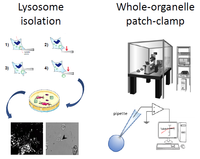
Image Credit: Image courtesy of Teresa Sorbo et al., in partnership with ELRIG (UK) Ltd.
LysoPatch on iCell® DopaNeurons
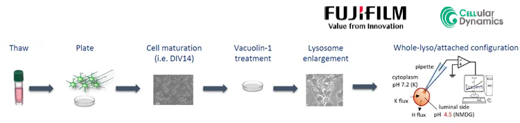
Image Credit: Image courtesy of Teresa Sorbo et al., in partnership with ELRIG (UK) Ltd.
Dopaminergic neurons carrying different TMEM175 mutations:
- AHN: Apparently Healthy Normal, M393T heterozygous (a.a. 393 M/T)
- M393M: homozygous (a.a. 393 M/M, engineered)
- T393T: homozygous LOF (a.a. 393 T/T, engineered)
- Q65P: heterozygous GOF (a.a. 393 M/M homozygous, a.a. 65 Q/P, engineered heterozygous)
TMEM175 overexpressing cell lines generation and clone validation
Cell line generation and clone selection using thallium response
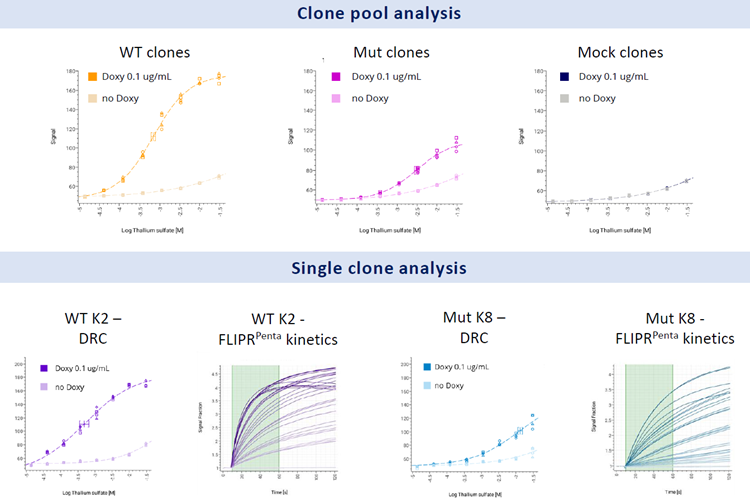
Image Credit: Image courtesy of Teresa Sorbo et al., in partnership with ELRIG (UK) Ltd.
- Both pools showed a good response to thallium sulphate.
- Only a slight activation was detected in the mock and not induced cells; the signal was significantly different from that of the induced clones, highlighting its specificity.
- Twelve well-performing clones were further characterized, and the top six responder clones from each cell line were selected and tested at SP384.
Clone selection at SP384
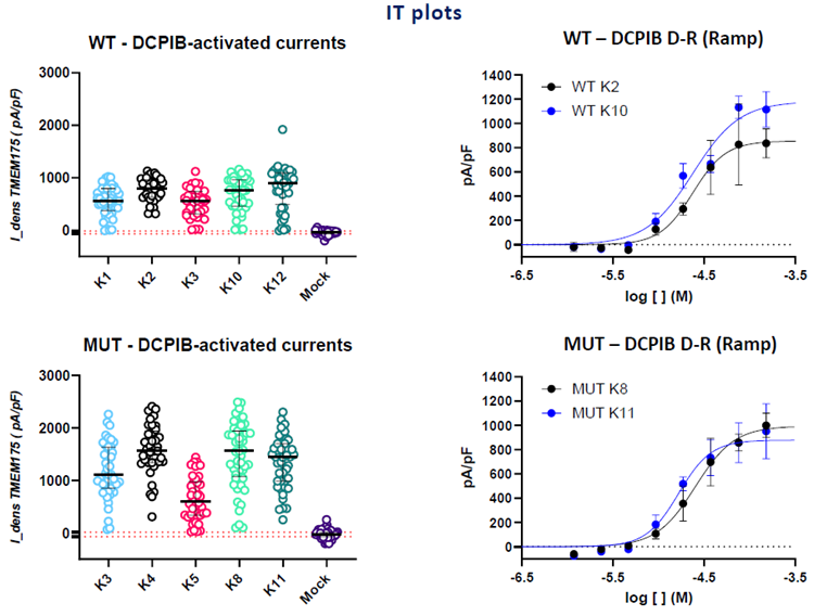
Image Credit: Image courtesy of Teresa Sorbo et al., in partnership with ELRIG (UK) Ltd.
- A voltage ramp protocol was used to evaluate the current amplitude at +100 mV.
- All tested clones responded to 100 μM DCPIB and were blocked by 2 mM 4-AP, achieving a good overall success rate (catch, seal, and expression).
- All the clones showed an outward current compatible with TMEM175 and statistically different from mock cells.
- WT K2 and K10, Mut K8 and K11 were chosen for further characterization.
Assay optimization at SP384
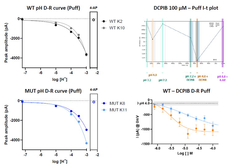
Image Credit: Image courtesy of Teresa Sorbo et al., in partnership with ELRIG (UK) Ltd.
- DRCs for DCPIB are reliable in all the investigated clones, and the EC50 values are comparable to those observed previously.
- A significant increase in peak current (proton flux) was recorded upon acidification using the puff method (Nanion).
- The DRC for DCPIB was also evaluated using the puff method at pH 7.2 and 4. The net proton current increases in response to increasing DCPIB concentrations.
TMEM175 at the lysosomal level
Automated patch clamp
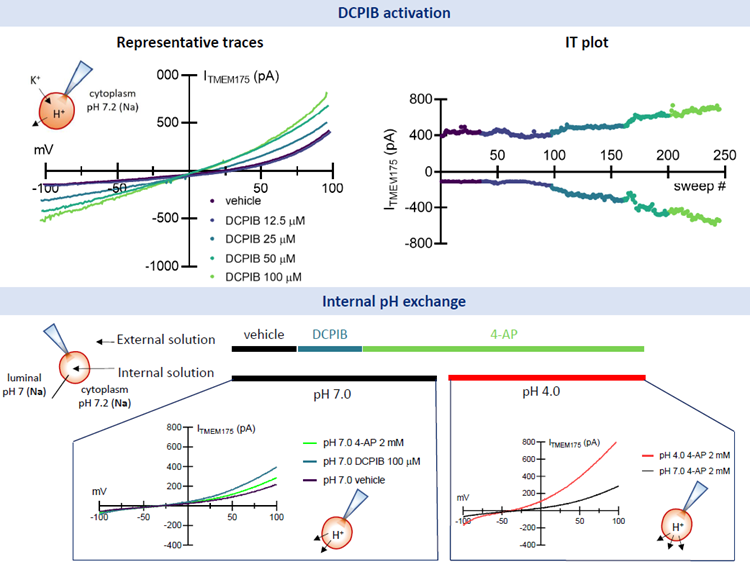
Image Credit: Image courtesy of Teresa Sorbo et al., in partnership with ELRIG (UK) Ltd.
- In-house protocol has been optimized to isolate high-quality lysosomes from the generated cell lines, and the material is suitable for automated LysoPatch experiments.
- DCPIB clearly activates TMEM175 currents at the lysosomal level, with an EC50 comparable to that observed in whole-cell recordings.
- Internal solution exchange enables the isolation of TMEM175 H+ and K+ conductance.
iPSC-derived dopaminergic neurons
Whole-lyso recording with DCPIB activation and single channel characterization in iCell® DopaNeurons
Whole-lyso configuration
- The DRCs to DCPIB are comparable in all conditions (cell line, current read-out) and similar to what has already been observed in HEK-cells.
- TMEM175 Q65P mutant line showed the highest activation, while the homozygous M393T Mutant showed, as expected, the lowest activation.
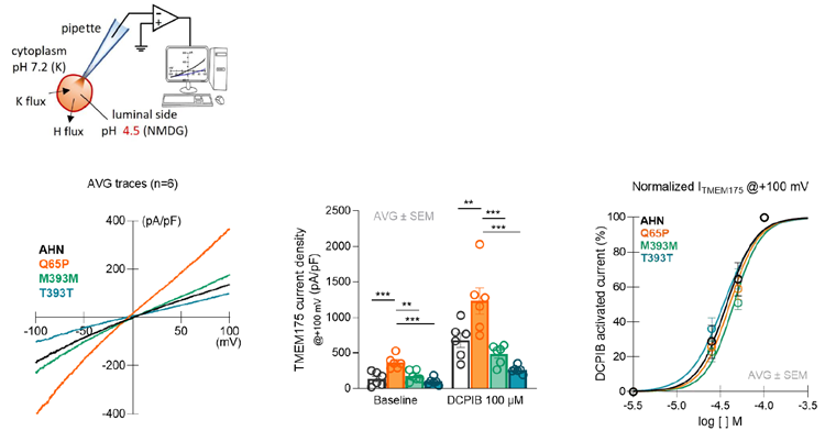
Image Credit: Image courtesy of Teresa Sorbo et al., in partnership with ELRIG (UK) Ltd.
- iCell® DopaNeurons with different TMEM175 mutations associated with Parkinson’s Disease and other α-synucleinophaties were cultured up to 14 days.
- The lysosomes of the four cell lines were patched manually in two different configurations.
- Preliminary data (n=5) on single-channel recordings indicate a higher number of channels in Q65P compared to the other cell lines, while open probability and dwelling time parameters are higher in M393M.
Lyso-attached configuration
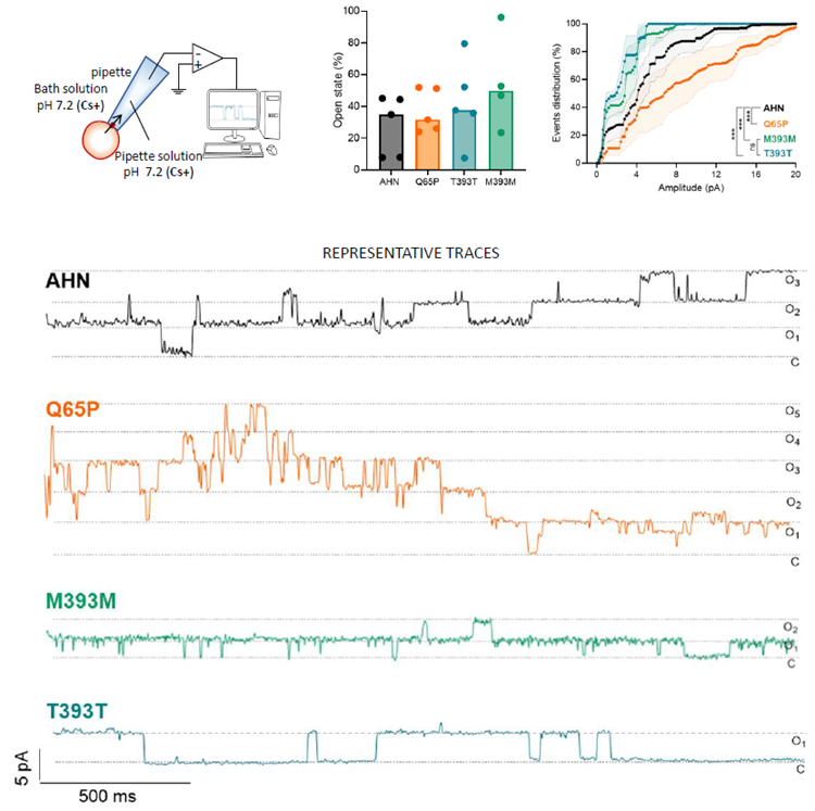
Image Credit: Image courtesy of Teresa Sorbo et al., in partnership with ELRIG (UK) Ltd.
Conclusions
- Stable cell lines expressing wild-type (WT) and mutated TMEM175 were successfully selected using a thallium fluorescence assay and further characterized electrophysiologically. All tested clones exhibited consistent responses to the TMEM175 activator DCPIB, acidic pH, and were effectively blocked by 4-AP, with a high success rate for catch, seal, and expression.
- Building on initial studies at the plasma membrane level, the team extended their investigations to the lysosomal membrane using both manual and automated patch-clamp techniques. These methodologies enabled precise measurement of TMEM175-mediated ionic currents within isolated lysosomes.
- Furthermore, lysosomal function was evaluated in iPSC-derived dopaminergic neurons (iCell® Dopaneurons, FCDI), providing a physiologically relevant model to explore TMEM175 in the context of neurodegenerative diseases. This approach provided a unique opportunity to integrate mechanistic insights into TMEM175 function using disease-relevant cellular models, further advancing our understanding of its role in lysosomal health and its potential as a therapeutic target for neurodegenerative conditions.
About Axxam S.p.A.
Axxam S.p.A. is a privately owned innovative Partner Research Organization (iPRO) and discovery company located at Openzone Science Park, Bresso (Milan, Italy), with chemistry laboratories located in Naples (Italy) and business development offices in Europe, US and Japan. Since its inception in 2001, Axxam has provided excellent quality discovery services to its clients, distributed around the globe and active in the Life Sciences as pharmaceuticals, crop protection, animal health, cosmetics, fragrances, food and beverages.
About ELRIG (UK) Ltd.
The European Laboratory Research & Innovation Group (ELRIG) is a leading European not-for-profit organization that exists to provide outstanding scientific content to the life science community. The foundation of the organization is based on the use and application of automation, robotics and instrumentation in life science laboratories, but over time, we have evolved to respond to the needs of biopharma by developing scientific programmes that focus on cutting-edge research areas that have the potential to revolutionize drug discovery.
Comprised of a global community of over 12,000 life science professionals, participating in our events, whether it be at one of our scientific conferences or one of our networking meetings, will enable any of our community to exchange information, within disciplines and across academic and biopharmaceutical organizations, on an open access basis, as all our events are free-of-charge to attend!
Our values
Our values are to always ensure the highest quality of content and that content will be made readily accessible to all, and that we will always be an inclusive organization, serving a diverse scientific network. In addition, ELRIG will always be a volunteer led organization, run by and for the life sciences community, on a not-for-profit basis.
Our purpose
ELRIG is a company whose purpose is to bring the life science and drug discovery communities together to learn, share, connect, innovate and collaborate, on an open access basis. We achieve this through the provision of world class conferences, networking events, webinars and digital content.
Sponsored Content Policy: News-Medical.net publishes articles and related content that may be derived from sources where we have existing commercial relationships, provided such content adds value to the core editorial ethos of News-Medical.Net which is to educate and inform site visitors interested in medical research, science, medical devices and treatments.
Last Updated: Nov 13, 2025