This article is based on a poster originally authored by Elena Baranova, Sabine Iglesias, Amanda Finan, Manon Motte, Renaud Burrer, Rania Gaspo, and Marie Gérus-Durand.
Limited tissue availability in immuno-oncology (I/O) emphasizes the need for precise characterization methods of the tumor microenvironment (TME). Extensive phenotyping of immune cell populations and the acquisition of unique biomarkers are crucial for understanding pro- and anti-tumor dynamics and for designing immunotherapy strategies. Multiplex immunohistochemistry (mIHC) has proved an effective way to achieve this.
Method
Panel development and staining platforms
Five mIHC panels were optimized by using two automated staining platforms: Leica Bond Rx and Roche Discovery Ultra.
Akoya Opals and Roche Chromogens were the detection chemistries used for fluorescence-based panels and chromogenic assays, respectively. Marker combinations were chosen based on their ability to quantify immune subsets, functional states, and tumor-immune interactions.
Tissue cohort
Panels were applied to normal and tumor tissues from the pancreas, lung, and colon. For each type of tissue:
- Five tumor samples
- Two healthy donor blocks

Figure 1. ROI Mapping on an ovary cancer sample. Image Credit: Cerba Research
ROI selection and mapping
Four regions of interest (ROIs) per block on hematoxylin- and eosin-stained (H&E-stained) slides were analyzed by a board-certified pathologist. To ensure consistent analysis across all samples, the ROIs were mapped precisely to the analogous multiplex panel slides.
Method: Image acquisition and analysis
Once images were captured, they were processed using the same internally validated workflow, which does not require spectral deconvolution. Quantification was conducted via HALO® (Indica Labs).
Metrics retrieved included:
- Percentage of positive cells
- Cell density (cells/mm2)
- Phenotypic combinations derived from co-expression patterns
For each sample, combined marker and phenotype levels were grouped across all ROIs. These values were distinguished from any whole-tissue measurements from the same slide. Matched-pair analysis was performed using JMP statistical analysis software, allowing direct comparison between ROI-based and whole-slide quantification.
Results: Multiplex IHC panels and associated phenotypes in a colon tumor sample
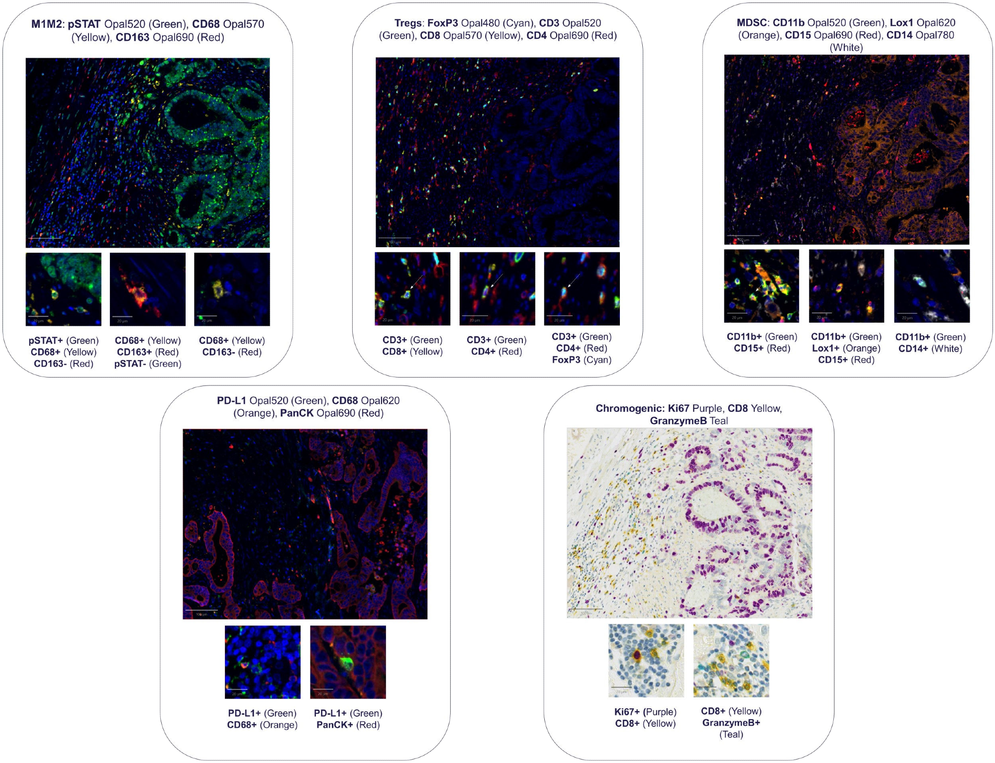
Figure 2. Multiplex IHC panel images are shown at the top of the layout, with the specific targets included for each panel labeled above the corresponding image. Beneath each multiplex panel, the associated phenotype images are presented, with the phenotype markers identified directly below each image. Image Credit: Cerba Research
Results: Image analysis results - Examples of visualizations
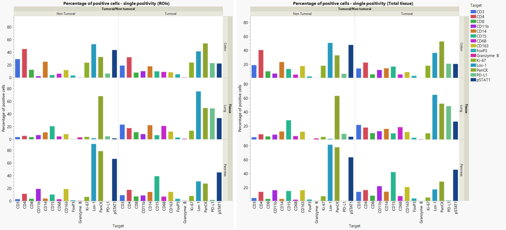
Figure 3. Quantification of single-positive cell percentage and cell density. Image Credit: Cerba Research
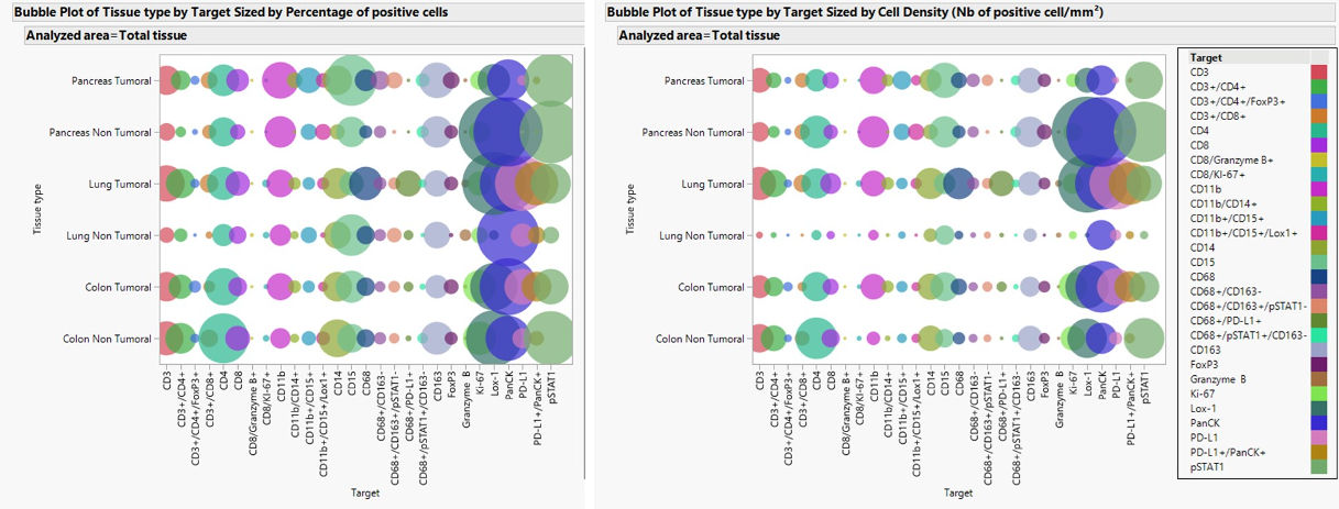
Figure 4. Visualization of the proportion of positive cells and cell density for single-marker positivity and phenotypes for each target/phenotype in total tissue. Image Credit: Cerba Research
Results: Total tissue vs ROIs comparison
Table 1. Total tissue vs ROIs comparison results. Source: Cerba Research
| Reportable parameter |
Target vs Phenotype |
Correlation |
| All |
All |
0.93 |
| Cell Density (Nb of positive cell/mm2) |
All |
0.93 |
| Phenotype |
0.95 |
| Single positivity |
0.91 |
| Percentage of positive cells |
All |
0.96 |
| Phenotype |
0.95 |
| Single positivity |
0.95 |
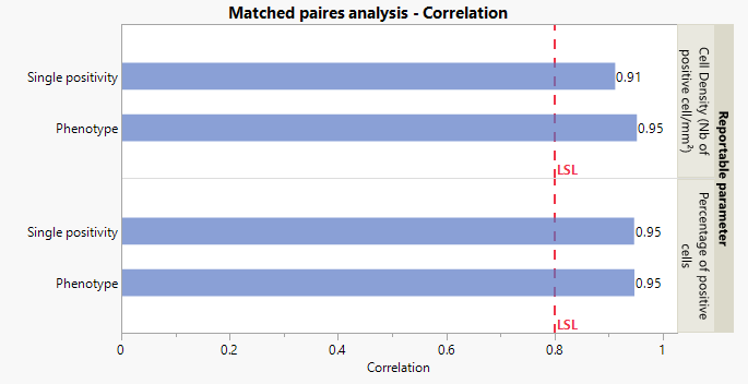
Figure 5. Matched pairs analysis using JMP software for percentage of positive cells and cell density for single positive cells and phenotypes. Image Credit: Cerba Research
Matched-pair analysis showed close agreement between ROI-based data and whole-tissue marker expression, indicating that the selected ROIs accurately represented the overall tissue context. This was applied to the following:
- Single-marker quantification
- Phenotypic combinations
- All reportable parameters
Multiplex panels demonstrated exceptional versatility across several tumor indications, showing a distinct differentiation between immune profiles in healthy and tumor tissues.
Detailed insights were obtained by quantifying single-marker positivity (e.g., CD8, CD68, PD-L1) alongside derived phenotypes (e.g., macrophage subsets, activated cytotoxic T-cells, regulatory T-cells), highlighting the spatial distribution and functional organization of immune subsets across the TME.
The study further underscored the following:
- Strengths of ROI-based analysis for focused, pathologist-driven evaluation
- Complementary value of whole-slide analysis for global tissue characterization
Conclusion
mIHC proved to be a powerful, dynamic, and instructive tool for immune profiling across solid tumors. By leveraging the combined utilities of automated staining platforms, high-resolution imaging, and quantitative image analysis, a detailed characterization of immune markers and phenotypes facilitated the acquisition of both cellular abundance and spatial context.
Through the incorporation of ROI-based and whole-slide quantification, this work enables a more comprehensive understanding of tumor immune interactions, facilitating cross-tissue and cross-tumor comparisons, as well as analysis of immune infiltration patterns and assessment of functional immune states within the TME.
These multiplex IHC workflows should be considered a robust analytical framework for the development of immuno-oncology biomarkers, while contributing key insights to advance precision immunotherapy.
About Cerba Research

Cerba Research is a leading specialty laboratory services provider with the capacity and breadth of a global central laboratory network. Their highly qualified scientists provide insight on the latest biomarkers, assays and testing approaches and develop innovative solutions for unique challenges across all research phases, to pharmaceutical, biotechnology, medical device, government, public health, and CRO organizations.
Cerba Research’s extensive capability in laboratory testing and global logistics including Bioanalysis, Flow Cytometry, Histopathology, and Next-Generation Sequencing, enables them to drive operational agility at scale in a wide range of therapeutic areas, with recognized expertise in Virology, Immunology, Oncology and Cell & Gene Therapy.
Cerba Research is part of the Cerba HealthCare Group with 15,000 employees on five continents, driven to advance diagnosis and health.
For more information about Cerba Research, please visit cerbaresearch.com.
Sponsored Content Policy: News-Medical.net publishes articles and related content that may be derived from sources where we have existing commercial relationships, provided such content adds value to the core editorial ethos of News-Medical.net, which is to educate and inform site visitors interested in medical research, science, medical devices and treatments.
Last Updated: Jul 16, 2026
 Cerba Research Expands U.S. Immuno-Oncology Center of Excellence to Strengthen Its Precision Medicine Offerings