Cell imaging techniques often rely on the use of dyes or genetic engineering to introduce fluorescent proteins.
Although it enhances contrast in the images, they can interfere with cellular activities. It can be hard to know whether these manipulations cause any fluctuations from the normal cell behavior. Ptychography presents a recently developed non-invasive method to study cells and cell cultures without the use of labels or artefacts.

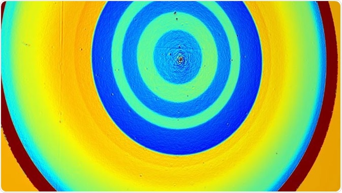
Phasefocus Virtual Lens® is a novel method for high fidelity quantitative imaging and microscopy. It is known in the scientific literature as "ptychography". Image Credit: Phasefocus
Method
There is an illumination beam shining on the specimen, behind which a camera is placed. Both the specimen and the illumination beam are moved in proportion to one another. This result in a series of partly overlapping illuminated areas on the specimen. The pattern of light diffracted by the specimen for each illuminated area is caught by a camera which is a charge-coupled device (CCD) camera. CCD facilitates conversion from pixels to digits or vice versa. The diffraction pattern image is further processed using a ptychography algorithm.
Phasefocus Virtual Lens
Advantages of Ptychography
Ideal for transparent samples
The diffraction pattern emerges from light has passed through the specimen, and this occurs with high accuracy and contrast. This makes it ideal for structural studies of nearly transparent cells. Ptychography is also less constrained in image resolution – the resolution is only limited by the effective numerical aperture (range of angles from which light can be detected by the lens) of the detector.
3D analysis
The image formation is not done by the detecting lens itself, but by the ptychography algorithm. An example of one such algorithm is the extended ptychography iterative engine (ePIE). ePIE algorithms produce two images: one of the amount of light that has been absorbed by the specimen and one of the amount of phase delay introduced to the illumination as it passes through the specimen. The depth of the images can be decided after acquisition. This is highly beneficial for 3D analysis of specimens and more complex cell cultures, a field which is continuously being developed.
Not restricted by optics of the system
ePIE also conducts calculations of the complex waveform of the illumination. Calculating the waveform allows for high quality reconstruction even when the illumination form is not known. This also avoids restrictions arising due to the optical system and places more emphasis on the algorithm. Several developments are therefore focusing on improving and extending the algorithm.
Life Sciences Applications
Increased brightness
Cells in the A549 cell line were analyzed using ptychography and compared to another microscopy technique called DIC (Diffrential Interference Contrast). The ptychography imaging is more similar to fluorescent imaging. It does not have a halo around the cells, as seen with other techniques. The areas inside the cells have brighter pixels than those outside the cell, making distinction easy. The nucleus can also be seen as the brightest point in the cell, a distinction which is harder to make with DIC microscopy.
Increased contrast
Certain cells had higher contrast than other in the study mentioned before. The authors hypothesize that this could be due to those cells entering the G2/M phase prior to mitosis. The main advantage of ptychography has been its ability to show distinctions between cells in different stages of the cell cycle. Other non-invasive or label-free imaging techniques have struggled with this. In addition, the full cell cycle of a A549 cell takes 18 hours. Ptychography was performed every five minutes for 20 hours, giving images of the full cell cycle for many cells. This allowed researchers to track and identify the changes happening.
Applications in Bacterial Imaging
Ptychography has also been applied to bacteria, without the need to isolate the samples. Deinococcus radiodurans has been in focus for its high tolerance towards ionizing radiation and possible correlations to its nucleoid structure.
Electron microscopy techniques applied have yielded contradicting results. Using ptychography, researchers found four distinct subunits present inside the bacteria which they identified as nucleoids owing to their general size and shape. The authors therefore believe ptychography can be used as a viable method to analyze bacterial nucleoids, independent of electron microscopy.
Further Reading
Last Updated: Aug 23, 2018