By Jeyashree Sundaram, MBA
Transmission electron microscopy (TEM) is a technique used to study the structure of small molecules like proteins or viruses, as well as other particles in material science.
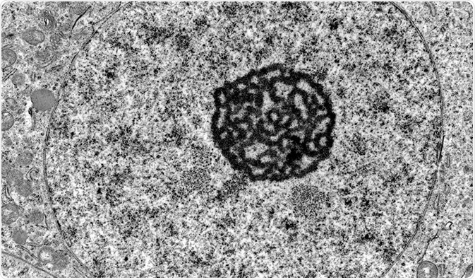
Credit: Jose Luis Calvo/Shutterstock.com
In this process, the particle to be studied is exposed to electron beams under a high-resolution microscope called a transmission electron microscope and the micrographs or images captured are analyzed computationally.
Sample preparation is a very crucial step in TEM and the method involved in preparing the sample differs, depending on the nature of the material and the information required from it.
Sample preparation in TEM
The process of specimen preparation in TEM involves many steps:
Fixation: Fixation of the specimen stabilizes the cell so that further change or damage to the cell will not happen. Through this process, the sample is preserved to give a snapshot in timeof the living cell. Fixation can be done through two methods as follows:
- Chemical fixation: This method is used for stabilizing biological samples. Chemical substances are used tocross link protein molecules with nearby molecules. The most frequently used chemical in this method is glutaraldehyde.
- Cryofixation: This method involves rapid freezing of the sample in either liquid nitrogen or liquid helium . The water content in the sample thus gets transformed into a vitreous ice form.
Rinsing: The tissue fixation process may cause increased acidity in the specimen. To prevent this condition and maintain the pH, it should be rinsed properly using a buffer such as sodium cacodylate.
Secondary fixation: To increase the contrast of the minute structures inside the specimen and give more stability, a secondary fixation is carried out using osmium tetroxide (OsO4). Without inducing any change in the features of the structure, OsO4 transforms the proteins into gels and increases the contrast between nearby cytoplasm by binding regions of phospholipid heads.
Dehydration: Freeze drying, or dehydration, of the specimen is the process by which the water content in the specimen is replaced with an organic solvent. Ethanol and acetone are the frequently used solvents in this method. Dehydration is important as the epoxy resin used in further steps does not mix with water.
Infiltration: In infiltration, epoxy resin is used to penetrate the cell, which will then occupy the space and make the sample hard enough to bear the pressure of sectioning or cutting. This process is also called embedding. The resin is then kept in an oven at 60° overnight to allow for setting. This process is called polymerization.
Polishing: After embedding, some materials are subjected to polishing. Polishing a specimen reduces scratches as well as other problems that can minimize the quality of the image. Ultrafine abrasives are used to give the specimen a mirror-like finish.
Cutting: For study under an electron microscope, the sample should be semi-transparent to allow the passage of electron beams through it. To achieve this semi-transparent nature, the sample is sectioned into fine sections using a glass or diamond knife attached to a device known as ultramicrotome. The device has a trough that is filled with distilled water.
The sections cut are collected in this trough and are then moved to a copper grid to be viewed under the microscope. The size of each section should be between 30 nm and 60 nm to get the best resolution.
Staining: Staining in biological specimens is usually done twice – before dehydration and after sectioning. In this process, heavy metals like uranium, lead, or tungsten are used to increase the contrast between different structures in the specimen, and also to scatter the electron beams.
Staining before hydration is done in block, while in staining after sectioning, the sample is exposed briefly to an aqueous solution of the above metals.
A cryofixed specimen may not undergo all these procedures. It can be directly subjected to cutting and then shadowed using vapors of platinum, gold, or carbon before visualization under the TEM.
Transmission Electron Microscopy
Credit: vulgarisation/Youtube.com
Apart from the above general procedures that are followed in preparing a sample for TEM, many other techniques are also available, such as:
- Ion-mining: in this process, thinning of the sample is done by firing charged argon ions at the sample surface until it becomes transparent enough. The focused ion-mining technique uses gallium ions for tinning.
- Cross-sectional method: This method is majorly used in the study of interfaces.
- Replica technique: Used only if the bulk specimen used to prepare thin sections cannot be damaged.
- Electrolyte polishing: This procedure is used to make thin samples out of metals or alloys. Coring, rolling, grinding, peeling, etc., are the different methods included in this procedure.
Further Reading
Last Updated: Jul 19, 2023
 New hybrid lens design drastically cuts 3D microscopy costs and complexity