An interview with Dr Claudia Jaffe, conducted at SfN by Alina Shrourou, BSc.
What are illuminators and how do they enhance microscopy and bioanalysis?
Light plays a critical role in our ability to generate high resolution images and to detect in a quantitative manner. In terms of image quality, it defines the contrast, resolution and signal to noise ratio. In quantitative measure, light defines the dynamic range, sensitivity and the lower detection limit. It ultimately plays a critical role in our ability to image and analyze.

© successo images/Shutterstock.com
Our illuminators are called light engines. A light engine is a family of light sources and all the optical elements, such as filters and mirrors, required to deliver the light to the sample. The intention is for it to work as simply as turning a lamp on and off. We can have as many as 50 to 70 optical elements in a single light engine, including more than a half dozen individual, independent light sources.
How is the light source or laser output in an illuminator related to the type of microscopy that can be performed?
We produce light sources for transmitted light, for absorption, and primarily for fluorescence. In each case, the quality of the light which is required is different. Fluorescence is the most demanding in the sense that it's not a particularly efficient process, so you have to put in billions of photons in order to get out enough to visualize something.
They have to be well constrained to certain spectral regions so that they overlap with the absorption bands of the fluorophores that are being interrogated. Tailoring the light to the spectral, spacial and temporal properties of the analysis instrument is quite challenging and a paradigm that we haven't had in technical lighting. In general, lasers or lamps produce certain spectra, brightness and angular output. Additional filters or mirrors are then required to direct light to a sample.
At Lumencor we use a more additive approach. If red/green/blue light is needed, we add only a red, a green and a blue light source. If you're illuminating a microtiter plate, a microscope slide, or a tiny spot of DNA, we focus the ray bundle to illuminate at angles and the area of interest. Similarly, regarding timing, lamps are usually on continuously. If you want to use them intermittently, you typically move a mechanical shutter in front of them.
We only use solid state lighting in our products, and that’s very stable. We can turn the light on and off in tens of microsecond time frames. For instance, in a very demanding application such as multicolor fluorescence live cell imaging, we’re trying to watch a biological process in real time.You can flash multiple colors on and off at 200 and 400 frames per second, and the light is only on when the camera is capturing data. That way, it's synchronized to the collection device which is on when needed but is off most of the time. This is a much more efficient use of photons.
Please give an overview of the light engines you highlighted at Neuroscience 2018.
In the case of neuroscience, we typically think about being able to switch light on and off for neurostimulation, and high brightness is often required. We're looking for outputs that stimulate the fluors that have been designed for those particular spectral regions, throughout the visible and into the near IR. We're looking for fast switching times and need to attenuate the amount of light delivered to a sample.
Often, the light is sent through a narrow bore fiber of 100 to 200-micron diameter, or smaller, to penetrate into a living brain or complicated neural structures within a brain slice, limiting the mechanical damage done to the tissues under study. We have light engines that do just that - generate white light, color lights, high brightness for each, outputs that are specifically designed to go into very small openings, like these narrow bore fibers, and extremely fast switching times.
As well, we are a very customer driven team. We look to cutting edge researchers to help us define what is needed for next generation experimentation. Our light engines are customizable and we often tailor the output for the specific user’s need.
What challenges in microscopy and bioanalysis do these light engines help researchers to overcome?
Their stability, longevity, and consistency is excellent because they are solid-state sources. That is very important for neurostimulation experiments because you want to be sure that all the artifacts that you observe are due to the biology and not imposed by the light. They run off a DC source, so unlike a lamp which most typically requires an AC driver that contributes its own noise, the DC source is quite flat. Also, solid-state sources don't have the familiar biphasic decay that lamps have, so our light output is quite flat and well behaved.
Brightness is often something that can limit your ability to detect or visualize something, and we tend to have best-in-case brightness in the life science marketplace. Traditionally, green light is very underserved by LEDs. The notorious “green gap” at 500 – 600 nm light is uniquely important in life sciences and ironically it’s the weakest output of most LEDs.
Lumencor uses a proprietary solution called a light pipe to overcome this optical power shortcoming. A narrow rod of doped glass or single crystalline material is excited either optically or electronically to generate light of these wavelengths. That light is essentially generated throughout the volume of the rod and trapped by total internal reflectance. The integrated output is released at a very small cross section at the end of the pipe.
This concentrated light output effectively boosts the intensity of the green and yellow light for Lumencor’s products. In many solid-state solutions where they promote an LED lamp, that spectral region is their least bright. In Lumencor's products, the green spectral region is often our brightest, making our products quite unique.
In terms of longevity, robustness, and environmental friendliness the light engines are green technologies. Unlike many arc lamps, we don't use any mercury in our products. Simon Watkins, Ph.D. at the University of Pittsburgh’s Center for Biologic Imaging is a technical adviser to the company and a heavy user of Lumencor’s products. He has at least a dozen light engines, purchased more than ten years ago when we first started manufacturing.
He often tells me I should promote not only the fact that they are still operating at ninety percent of their initial output power, but the cost savings associated with no replacement parts. He needs no replacement bulbs, light guides or filters, and so saves thousands of dollars in disposables each year. There is a slow, steady decay in signal over a long time but no catastrophic failure, and so 10,000 to 20,000 hours of quality output is common for these illumination tools.
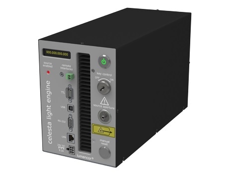
Can you talk about any examples where the CELESTA light engine has been used in optogenetics research?
Celesta is unique in that it is a multiline laser light engine. It’s our only product that is driven exclusively by solid state lasers. Celesta is designed for high brightness applications, like confocal microscopy, TIRF, super-resolution microscopy, optogenetics, FRAP and SPIM. These are applications where you really need a lot of light and/or a lot of brightness. So many tens or hundreds or even thousands of mWs of power and/or lots of power out a very narrow bore fiber. We can provide factors of 3 - 5x more brightness than the predecessor products from competitive companies.
Often we are competing with multiline laser systems that offer half the number of outputs (three or four lines vs. seven colors in Celesta) at more than twice the price. Moreover, laser users are often manipulating many components on an expensive laser table that takes up signicant laboratory real estate. Our box is plug-in ready, occupies a small tabletop footprint and requires no alignment or mainentance. We see it as a game changer.
What do you hope to gain from Neuroscience 2018?
SfN is the place equipment manufacturers come and feature their most exciting and newest technology. I saw some fantastic computational clearing technology in the Leica booth. They are using intelligent algorithms to clean up optical cross sections of complicated multicolor samples. They can eliminate blur and take out background signal and create crystal clear images. You see that first at Neuroscience. We’re very pleased to be a long time supporter of this meeting and the fabulous work those in attendance contribute.
About Claudia B. Jaffe, PhD
_Lumencor.jpg)
Claudia Jaffe earned her doctorate in Bioanalytical Chemistry from the University of Pittsburgh. She has developed, published and patented a variety of electrochemical and photoelectrochemical sensors and bioanalytical chips, focusing her efforts on high throughput analyses employing enzymology, immunology and genomics. She was an initial employee of Caliper Technologies and generated the first families of fluorogenic and separation based assays for compound library screening in lab-on-a-chip devices.
At Protogene Labs, she developed applications in Q-PCR utilizing novel ink jet printed, customizable microarrays, now produced at Agilent Technologies. At Lumencor, she is an inventor in nearly all the company’s patents and the leader of new business development. As well, she supervises sales and marketing, generating key new business relationships in developing markets for Lumencor’s novel lighting products in the life science arena.