In this interview, News Medical speaks with Anna Paszulewicz, Product Manager for Imaris at Oxford Instruments, about Imaris 11.1 and its latest developments in microscopy image analysis. She discusses how the new Imaris Training Hub helps researchers access tutorials, validated workflows, and educational datasets directly within the software. She also explores how improvements to data handling, workflow transparency, and access to training resources can help researchers work more efficiently, develop their image analysis skills, and obtain more consistent results from complex microscopy datasets.
Can you please introduce yourself and your role at Imaris?
My name is Anna Paszulewicz, and I am the Product Manager for Imaris at Oxford Instruments. I work on developing the Imaris platform to help researchers get more from their microscopy data and improve their image analysis workflows.
My focus is on understanding the challenges researchers encounter when analyzing increasingly complex datasets and developing software capabilities that make image analysis more accessible, efficient, and reliable. With Imaris 11.1, we are continuing this work by bringing learning resources directly into the software, helping users develop their skills and access practical image analysis examples without leaving their working environment.
What is Imaris 11.1, and what are the main objectives behind this release?
Imaris is a modular image visualization and analysis software platform that supports researchers throughout the stages following image acquisition, from data preparation and image analysis to visualization, quantification, and presentation of results.
With Imaris 11.1, we are placing particular emphasis on helping users learn more efficiently and work with greater confidence. The new Imaris Training Hub brings educational materials, tutorials, example datasets, and predefined workflows into the software environment.
We want to make it easier for researchers to understand how to approach an image analysis task, learn established methods, and apply them to their own data. By improving access to training and practical guidance, we aim to reduce the time users spend learning the software and help them focus more on their research questions.
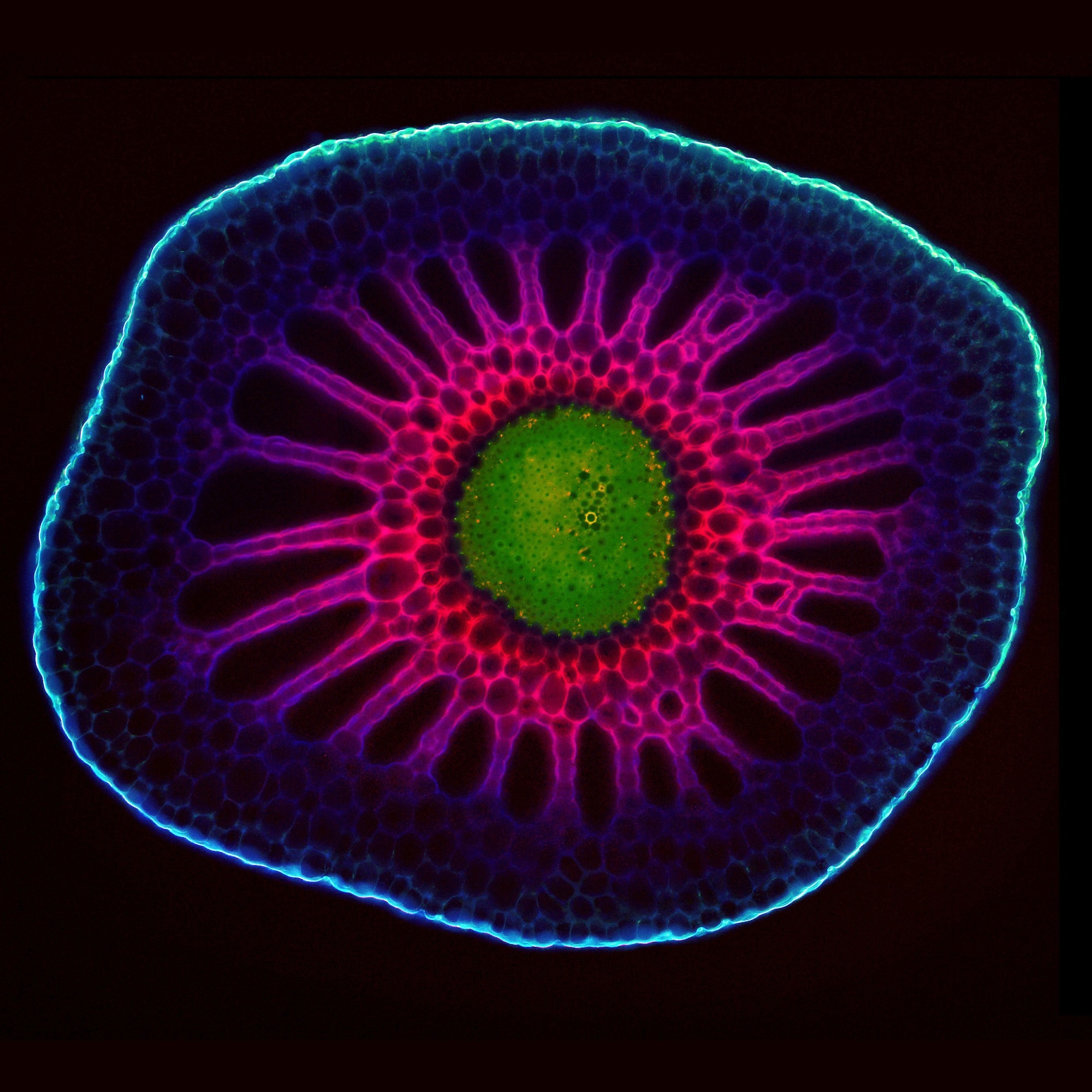 Source: Oxford Instruments - Life Sciences
What is the Imaris Training Hub, and how can it support researchers?
The Imaris Training Hub provides access to learning resources directly within Imaris. It brings together tutorials, training videos, example datasets and workflows that have been prepared and validated by our technical and applications teams.
This is particularly useful for researchers who are new to the software or who want to learn a new analysis technique. Rather than searching for information across different resources, users can access guidance from within the application itself.
The hub also helps users learn from a consistent source. By providing structured tutorials and practical examples, we can support the adoption of sound analytical practices and make it easier for researchers to understand how specific methods should be applied.
For experienced users, the resources can also provide a useful starting point for exploring additional analysis capabilities.
How does Imaris 11.1 help improve the reproducibility of image analysis?
Reproducibility has been an important focus of recent Imaris development. Image analysis can involve many individual steps, including image processing, segmentation, object detection, and quantification; when these steps are performed manually, it can be difficult to remember every parameter or repeat the same procedure consistently across multiple datasets.
Imaris introduced Workflows to capture the steps involved in an analysis protocol, allowing users to save and reuse their procedures. This helps reduce repetitive manual work and makes it easier to apply the same approach to comparable datasets.
Imaris 11.1 complements these capabilities by improving access to validated example workflows and training materials. Researchers can explore established approaches, understand how an analysis was performed, and use those examples to develop their own methods. Together, these capabilities help support more consistent image analysis across experiments and users.
Modular image visualization and analysis with Imaris 11.1 | Webinar Highlights: Oxford Instruments
How can researchers use the training resources to learn new image analysis techniques?
The key advantage is that users can learn within the same environment as where they perform their analysis. The Training Hub provides access to educational datasets, video tutorials, supporting documentation, and predefined workflows.
Researchers can use these materials to become familiar with the software, explore different analysis approaches, and practice using example data. They can also examine workflows to understand the sequence of operations used to achieve particular results.
This practical approach is valuable because learning image analysis is not simply about knowing which tools are available. Researchers also need to understand how to select appropriate methods and apply them to their specific datasets.
Having these resources readily available makes it easier to explore new techniques independently and build confidence before applying them to research data.
What improvements have been made to data management and navigation in Imaris 11.1?
Imaris 11.1 introduces improvements to Imaris Arena, helping users find and organize their files more efficiently. New search and sorting functionalities make it easier to navigate datasets and locate relevant materials.
These improvements are particularly useful when working with collections of microscopy data and educational resources. As projects develop, researchers may accumulate raw images, processed datasets, videos, documentation, and analysis workflows. Being able to locate the right material quickly can simplify the overall analysis process.
The release also introduces native support for ZEISS CZI files through an integrated reader. This helps streamline the import and conversion of these datasets within the Imaris environment.
These changes focus on reducing unnecessary steps in everyday work, allowing researchers to spend less time navigating files and more time analyzing and interpreting their results.
How does Imaris Viewer support collaboration and the sharing of image analysis results?
Imaris Viewer provides a way to open and explore microscopy datasets without requiring the full Imaris analysis environment. Researchers can use it to visualize raw microscopy data and examine datasets that have already been analyzed.
In Imaris 11.1, Viewer also allows users to inspect workflows associated with previously analyzed datasets. They can see the parameters used in each workflow step and review the image preprocessing applied.
This additional transparency is useful when sharing results with colleagues, presenting data at conferences, or discussing an analysis with collaborators. It provides more context about how the results were generated, rather than showing only the final visualization.
The workflow inspection functionality is read-only, so users cannot modify or export the workflow through Viewer. Nevertheless, it offers a valuable way to understand and review the analysis behind a dataset.
How can these developments benefit imaging facilities and research teams working with complex datasets?
Imaging facilities often support researchers with varying levels of experience, from those just beginning to work with microscopy data to specialists conducting advanced quantitative analysis.
Providing individual training and answering recurring questions can require considerable time. The Training Hub helps address this challenge by making structured learning materials and validated examples accessible directly within the software. Researchers can use these resources to learn common tasks and develop greater independence.
For research teams, shared training materials and workflows can also help establish more consistent approaches to analysis. This is especially relevant when multiple researchers are working on related experiments or when datasets need to be compared across experimental conditions.
By combining accessible training with workflow-based analysis, Imaris 11.1 aims to help facilities use their support resources more effectively while enabling researchers to work more confidently with their data.
How does Imaris 11.1 fit into the wider Imaris software ecosystem?
Imaris is part of a broader software family designed to support different stages of the microscopy workflow. Depending on their requirements, researchers can use tools for file conversion, large-scale image stitching, image analysis, visualization, and the presentation of results.
Imaris itself supports data preparation, analysis, exploration and presentation, including the creation of snapshots and animations. Researchers can also use Imaris Vantage to explore and visualize analysis results with plots, while the Batch module automates operations across multiple datasets.
These capabilities are important because image analysis requirements vary considerably between research projects. Some users may be working with a single dataset, while others need to process large numbers of images or compare results across experiments.
The continued development of these tools, alongside the new Training Hub in Imaris 11.1, reflects our aim to help researchers manage their data more effectively and obtain meaningful insights from their microscopy experiments.
About Anna Paszulewicz

Anna Paszulewicz holds a degree in Biophysics from Jagiellonian University in Kraków, Poland. Passionate about image analysis and microscopy, she has been dedicated to the Imaris platform for more than 13 years, working closely with researchers, imaging facilities, and development teams to translate scientific challenges into practical image analysis solutions. As Product Manager for Imaris, Anna focuses on helping scientists extract meaningful biological insights from increasingly complex imaging datasets.
About Oxford Instruments - Life Sciences
Oxford Instruments is a leading provider of high-technology tools and systems for research and industry, dedicated to accelerating breakthroughs that create a brighter future for our world. With a global presence, we are committed to innovation and excellence, offering cutting-edge solutions that enable researchers and industry professionals to achieve breakthroughs in their fields. Our advanced technologies deliver numerous benefits through unparalleled precision and reliability, allowing users to obtain accurate and reproducible results. By utilizing Oxford Instruments' innovative solutions, research is accelerated, productivity is enhanced, and innovation is achieved in various fields, including materials analysis, life sciences, semiconductors, physics, chemistry, and food sciences. We take pride in being a trusted partner for those aiming to push the boundaries of scientific and industrial advancements, providing the tools and support necessary to realize their visions.
Atomic force microscopy: These advanced instruments are utilized for high-resolution imaging and precise measurement of surface properties at the nanoscale level. They offer detailed topographical information and enable accurate measurements of features such as height, roughness, and mechanical properties.
Light microscopy: Our solutions encompass a comprehensive range of advanced imaging systems that utilize visible light for examining samples at the microscale. Equipped with high-quality lenses, cameras, and illumination systems, these optical microscopes deliver detailed images with exceptional clarity and resolution. They find applications in fields such as biology, materials science, and forensics.
Plasma etch & deposition: innovative etch & deposition techniques, atomic layer etch and atomic layer deposition, and ionbeam etch & deposition solutions used to produce high yielding, efficient and reliable advanced semiconductor devices. Our service and product range includes research and development systems right through to fully clusterable, robust and high up-time production systems, designed for high-volume manufacturing in key markets like quantum, power devices, augmented reality and datacom.
Electron microscopy analysis: Our high-performance tools are tailored for materials characterization, particle analysis, and sample manipulation at the nanometer scale. Techniques such as Backscatter Electron and X-ray (BEX), Energy Dispersive Spectroscopy (EDS), and Electron Backscatter Diffraction (EBSD) enable imaging, chemical analysis, and crystallographic characterization of materials at atomic and nanoscale levels.
Optical imaging and spectroscopy: Our optical imaging solutions comprise a range of advanced technologies and systems for capturing and analyzing images using light. From state-of-the-art optical microscopes to spectroscopy systems and imaging software, these solutions provide high-quality images and data for applications such as materials characterization, biological research, and semiconductor analysis.
Nanoindentation: Our high-resolution, MEMS-based nanoindenters are designed to measure the mechanical properties of materials, including hardness, elastic modulus, stiffness, and creep behavior. These instruments offer superior fabrication tolerances, enhancing sensitivity, resolution, and repeatability beyond conventional technology limits.
Nuclear Magnetic Resonance: We offer a variety of benchtop Nuclear Magnetic Resonance (NMR) instruments for research, industrial quality assurance/control, and rock core analysis. These instruments provide advanced capabilities for chemical analysis, materials characterization, and more.
Raman microscopy: Our advanced imaging systems are tailored for high-resolution microscopy and analysis, offering cutting-edge technology and precision optics for detailed insights into samples at the micro- and nanoscales. Trusted by researchers and scientists in fields such as materials science, life sciences, and nanotechnology, these systems are available in various models to suit different applications.
X-ray technologies: Our technologies encompass a range of advanced systems and solutions for materials analysis and characterization. With cutting-edge capabilities and high sensitivity, Oxford Instruments X-ray technologies are widely used in various industries and research academic institutions for applications such as material identification, quality control, and research in fields like geology, metallurgy, and semiconductor manufacturing.
 Imaris 11 transforms image analysis with automated workflows and reproducible research tools