Oct 23 2018
The discovery of mycoplasma contaminants in cell culture and the subsequent biologicals is vital because of the negatives effects mycoplasma has on the physiology of cells.
The contamination of mycoplasma in continuous animal cell cultures has been known to be a big issue for a long time, due to the broad range of effects that mycoplasma has on cells in culture. These effects range from modifications of cellular gene expression to activation of the signal transduction pathway (1,2), and lead to potentially compromised data due to the unintended effects of mycoplasma contamination.
Mycoplasma contamination in biopharmaceuticals is linked to the raw materials, cell lines and cell cultures that are used in production. Detecting contamination early on enables quicker choices to be made and corrective steps to be taken to conserve subsequent product integrity.
Commercial suppliers of cell lines need to be able to rapidly analyze cell cultures to detect microbial and, specifically, Mycoplasma contamination. An important aspect is keeping track of Mycoplasma contamination and assuring that cell lines are not contaminated.
Analysis of Mycoplasma is made easier with the new features from UVP GelStudio, which include automated out-of-the-box capture of fluorescent PCR gel results by one click. The automation feature extends to gel image annotation and allows the raw gel images to be labeled and archived efficiently and effectively.
Materials and Methods
Sample Preparation
10µl of cell culture supernatant was boiled for 10 minutes and briefly centrifuged. After which, 2µl of supernatant was added to rehydrated PCR master reaction mixture, which was followed by PCR. Agarose gel electrophoresis was then run, and the PCR product was analyzed by utilizing the bioimaging UVP GelStudio.
Samples and Reagents
- PCR was run with the PCR mycoplasma test kit (PK-CA91-1024 from Promokine)
- The kit contains primers, nucleotides, DNAs and hot start Taq polymerase required for PCR
- Rehydration buffer
- Positive controls
- Agarose gel electrophoresis
Instrumentation
Customizable action buttons are used by the UVP GelStudio to automatically focus, capture, pseudo-color and save images. Electrophoretically-separated DNA can be identified and quantified using automated tools, and routine gel annotations can be saved and loaded for each gel, which makes labeling and documentation easy.
The UVP GelStudio has a lowlight optical zoom lens, seen in Figure 1.2, which allows a broad range of gel sizes to be analyzed using preset one-click zoom settings. An extensive range of fluorescent dyes can be imaged, and new dyes or multiplexing can be accommodated for via the easy exchange of fluorescent emission filters. Furthermore, 21 CFR part 11 is supported by certain features.
For experimentation, the camera, focus, zoom, exposure, and excitation and emission filters can be manually controlled.
Results and Discussion
An extremely quick and sensitive work studio is provided by the UVP work studio for imaging and analysis of contamination by mycoplasma. Human and mouse tumor cells that express GFP and RFP fluorescent proteins were analyzed for mycoplasma presence using agarose gel electrophoresis.
A distinct band at 265-278 bp was seen in samples containing mycoplasma positive, whereas an internal control band and a negative control at 479 bp was seen in samples negative for mycoplasma contamination. Mycoplasma contamination was positive for LLC cells producing RFP fluorescent proteins and CT-26 cells producing GFP fluorescent proteins. Distinct bands show the internal control DNA on the gel, thus showing that the PCR was run successfully, and the data obtained is not erroneous (Figure 1.3).
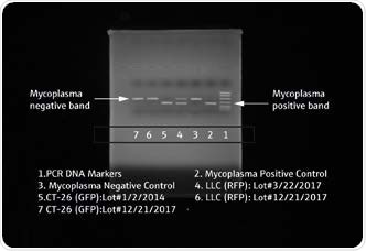
Figure 1.3. Agarose Gel of Mycoplasma PCR Test Results - Bioimaging results of agarose gel electrophoresis of PCR products from human and mouse tumor cells that express GFP and RFP fluorescent proteins. LLC (RFP) and C-26 (GFP) have the same number of base pairs as the Mycoplasma positive band DNA.
Promokine’s PK-CA91-1024 PCR mycoplasma kit does not identify clinically relevant mycoplasma species such as M. pneumonia and U. urelyticum but does detect species such as M. bovis and M. arthritidis.
Conclusion
Detection of mycoplasma at low concentrations in cell cultures is now possible using robust new methods. The UVP GelStudio’s innovative design allows it to fit effortlessly in the laboratory workflow, and it is easy and accurate when identifying and quantifying mycoplasma in cell cultures. Fluorescence DNA on a gel can be imaged and that raw image can now be labeled and analyzed efficiently. Data gained from bioimaging permits simple documentation and, furthermore, can aid in archiving and referencing future results from electrophoresis. Data loss will be reduced and data analysis will be facilitated due to the capabilities of the studio.
References
- Drexler, H.G. and Uphoff, C.C., 2002. Mycoplasma contamination of cell cultures: Incidence, sources, effects, detection, elimination, prevention. Cytotechnology, 39(2), pp.75-90.
- Stanbridge, E.R.I.C., 1971. Mycoplasmas and cell cultures. Bacteriological reviews, 35(2), p.206.
- Chen, T.R., 1977. In situ detection of mycoplasma contamination in cell cultures by fluorescent Hoechst 33258 stain. Experimental cell research, 104(2), pp.255-262.
Analytik Jena AG
Analytik Jena is a provider of instruments and products in the areas of analytical measuring technology, life science, and optoelectronics. Its portfolio includes the most modern analytical technology, complete systems for bioanalytical applications in the life science area, and high-end optical consumer product.
Sponsored Content Policy: News-Medical.net publishes articles and related content that may be derived from sources where we have existing commercial relationships, provided such content adds value to the core editorial ethos of News-Medical.Net which is to educate and inform site visitors interested in medical research, science, medical devices and treatments.