For years, the standard method for imaging the development of vertebrate skeletons has been the use of Alizarin Red and Alcian Blue for clearing and staining.1 These techniques have also been applied in the study of whole-embryo skeletal development.2
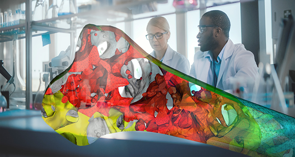
Image Credit: Thermo Fisher Scientific
By employing staining, researchers can obtain images with extensive anatomical detail that allows them to characterize animal models and identify developmental malformations. However, so far, most of this work has been limited to 2D imaging, restricting the level of detail achievable.
The introduction of novel approaches enabling 3D visualization of bone and cartilage presents a groundbreaking opportunity to enhance the understanding of vertebrate development.1,2
By transitioning from the traditional 2D approach to 3D imaging, researchers can unlock a new realm of information, leading to a deeper comprehension of the underlying processes that drive vertebrate skeletal development.
This paradigm shift offers exciting possibilities for the study of developmental biology, comparative anatomy, and the discovery of novel markers for developmental disorders.
Refining the process
The process has been refined through the utilization of optical projection tomography, which effectively 3D-images Alcian Blue stained cartilage in cleared specimens.3
Additionally, various modalities such as microcomputed tomography (microCT), 3D phase-contrast X-Ray tomography, and MRI have been explored for imaging purposes.3 MicroCT has enabled the 3D imaging of developing bone. This means that genuine 3D data could be collected at micron resolution.
Currently, there is no standardized workflow for reliable separation and intensity-based visualization of skeletal tissues, allowing simultaneous isotropic 3D imaging of cartilage and bone with similar quality.
To address this, a research team led by Stephan Handschuh at the University of Veterinary Medicine has endeavored to develop an efficient X-Ray microCT imaging protocol.4
Their innovative approach involves selectively labeling the cartilage matrix, providing adequate contrast compared to other tissues for automated visualization and segmentation while preserving the bone mineral. This enables simultaneous imaging of both tissues in the developing skeleton.
To establish the outlined protocol, the researchers conducted experiments, testing various fixatives, washing agents, and staining solutions.
After multiple trials involving different fixatives and contrast agents, they successfully developed a staining protocol utilizing Ruthenium Red in 50% ethanol for ethanol-fixed samples. Subsequently, the researchers put their protocol to the test using E16.5 mouse fetuses.
Drawing distinctions
The researchers quickly discovered that their protocol yielded detailed anatomical information about the skeleton, particularly focusing on crucial areas for studying skeletal development, such as the limbs and ribcage.
They successfully obtained morphometric measurements for these skeletal components. However, a challenge they faced was the overlap in X-Ray attenuation between fetal bone and cartilage.
To address this issue, the researchers devised a dual-energy protocol based on the distinct X-Ray attenuation properties of ruthenium and hydroxyapatite at two energy spectra. This innovative approach allowed them to reliably differentiate bone from cartilage.
By extracting material fractions with well-separated ruthenium and hydroxyapatite signals, the researchers generated 3D renderings that closely resembled specimens obtained through clearing and staining.
The advantage of this new method was the authentic isotropic 3D data it provided, enabling accurate morphometric measurements. It is also important to note that the protocol successfully separated the cartilage matrix from most organs and soft tissue.
This achievement marked the first instance of a high-contrast staining protocol that facilitated both intensity-based segmentation and visual phenotyping of cartilage elements, which can be used for quantitative and qualitative analyses of the cartilage developmental process.1
A promising alternative
Due to the simple and reproducible nature of the staining protocol, in the future, this method could be used for fully automated imaging and quantitative morphometric analysis, and high-throughput analyses. While the protocol is viable for use with lab-based scanners, additional hardware developments could help improve image quality.
The valuable information obtainable from authentic 3D data greatly benefits the study of skeletal development, with potential applications in developmental disorder screening and quantitative characterization of developmental malformations.
For instance, this approach facilitates the detection of abnormalities in limbs and ribcages, frequently studied in genetic disease models or developmental toxicity assessment of test agents.
The research team's innovative workflow presents a compelling alternative to conventional methods for vertebrate skeletal development phenotyping, proving indispensable across various sectors of the life sciences industry.1
Thermo Fisher Scientific technology
In this research, the team utilized Thermo Fisher Scientific technology, specifically Thermo Scientific Amira Software, to import and analyze the datasets for 3D reconstruction and analysis.
Amira Software provides a robust platform for visualizing, analyzing, and processing 2D-5D images across various imaging modalities. To support their study, the researchers utilized built-in tools such as the Amira Software volume segmentation tool Volume edit, and Surface Area Volume tool.
Amira Software offers both flexibility and speed, catering to advanced imaging workflows in numerous research areas, including structural and cellular biology, tissue imaging, bioengineering, and preclinical imaging.
Its powerful segmentation and image processing capabilities enable researchers to gain crucial insights from their data.5
References
- Hilton, M. J. (2014). Skeletal Development and Repair. https://doi.org/10.1007/978-1-62703-989-5
- Yamazaki, Y., Yuguchi, M., Kubota, S., Isokawa, K. (2011). Whole-mount bone and cartilage staining of chick embryos with minimal decalcification Biotechnic and Histochemistry, 86(5), 351–358. https://doi.org/10.3109/10520295.2010.506158
- Akhter, M. P., & Recker, R. R. (2021). High resolution imaging in bone tissue research-review. Bone, 143(August 2020), 115620. https://doi.org/10.1016/j.bone.2020.115620
- Gabner S, et al. The visible skeleton 2.0: phenotyping of cartilage and bone in fixed vertebrate embryos and foetuses based on X-ray microCT. Development 2020;147:dev187633. doi:10.1242/dev.187633.
- Amira software for Cell Biology (no date) Thermo Fisher Scientific - US. Available at: https://www.thermofisher.com/in/en/home/electron-microscopy/products/software-em-3d-vis/amira-software/cell-biology.html
Familiarize yourself with Amira Software for bone and tissue image analysis
With its advanced image processing, segmentation, quantification, and reporting features powered by artificial intelligence (AI), Amira Software can provide you with the tools you need to take your discoveries to the next level.
In this webinar, you will discover how it was used to:
- Easily identify and analyze structural bone regions and precisely examine and measure specific bone structures to gain a deeper understanding of bone health, growth patterns, diseases, or responses to various treatments
- Perform advanced characterization of the growth plates in murine knee bones to obtain valuable insights into skeletal development, growth disorders, and potential treatments for related conditions
- Obtain quantitative information of bone tissue microstructure to understand bone biomechanics and the impact of different factors on bone structure and function
Watch on-demand Webinar
Sponsored Content Policy: News-Medical.net publishes articles and related content that may be derived from sources where we have existing commercial relationships, provided such content adds value to the core editorial ethos of News-Medical.Net which is to educate and inform site visitors interested in medical research, science, medical devices and treatments.