In the context of examining bone health in pre-clinical models of diverse diseases, microCT proves highly valuable, particularly when it comes to imaging bones and conducting longitudinal quantitative assessments.1
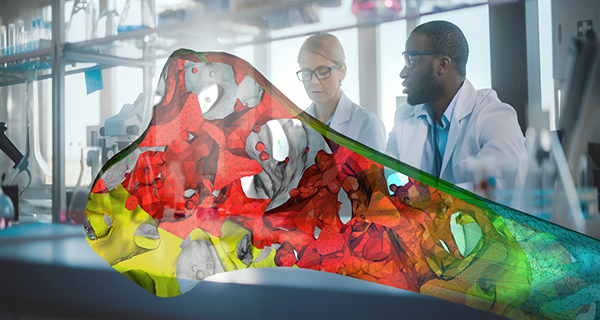
Image Credit: Thermo Fisher Scientific
Nonetheless, data analysis of micro CT imaging is limited in sample size due to the significant effort required for segmentation [2].
Presently, segmenting individual bones is generally reliant on manual contouring or density-based thresholding methods.
Due to potential inaccuracies and inter-user variability associated with both manual contouring and density-based thresholding methods, researchers may limit their analysis to just one bone or a subset of bones. This restriction consequently reduces the ability to identify bone health biomarkers for further investigation.
In response to these limitations, a team of researchers led by Hani Awad at the University of Rochester Medical Center, United States, has devised a high-throughput, semi-automated segmentation method for murine hindpaw microCT datasets.3
Developing a workflow using Amira Software
To establish an effective workflow, the research team utilized microCT datasets obtained from a large number of hindpaws belonging to mice at monthly intervals, ranging from two to eight months old for males and two to five months old for females.
Subsequently, the acquired data was exported to Thermo Scientific™ Amira™ Software. Within the software, the researchers employed watershed-based algorithms to carry out automated segmentation, which was then subject to user correction.
The process involved the utilization of filtered data and a binary mask as inputs, ultimately generating a fully segmented dataset.
The automated portion of the watershed seed placement exhibited an overall accuracy of 79.2% for in vivo bone segmentation. It is worth noting that error rates varied among different bones, with some displaying higher error rates than others. The majority of bones were accurately segmented without requiring user correction.
To address and rectify these errors, the research team devised effective methods. Upon the completion of the segmentation process, each bone volume was extracted and organized as a data table within Amira Software.
No experience required
The researchers conducted a comprehensive test of their workflow, involving one experienced user and three novice users who were unfamiliar with Amira Software. Novice users underwent training through instructional videos and practice data sets before reanalyzing the data, with segmentation time recorded.
The results demonstrated a strong correlation between the semi-automated method's outcomes and those achieved through conventional analysis by an experienced individual.
The study's results demonstrated a high level of internal consistency when the experienced user repeated the semi-automated segmentation for a second time. Additionally, the semi-automated workflow exhibited a significant time advantage over the conventional method.
Overall, the main findings revealed that the experienced user completed the segmentation process at an accelerated pace with the assistance of automation, achieving a speed nearly 10 times faster than the conventional approach.
The study further found that novice users could also complete their tasks much faster using the novel semi-automated approach compared to experienced users employing the conventional method. Notably, similar excellent inter-user reliability was observed for both novice and experienced users.
As a result, it can be confidently concluded that even a novice user with no prior experience with Amira could accomplish data segmentation after only 1–2 days of practice. Soon after this brief learning period, the user would be proficient in performing high-throughput and reliable data analysis.3
A promising approach for accelerated performance
By adopting the semi-automated method described in this study, the establishment of a standardized protocol for bone volume measurements across different users and institutions becomes feasible.
The high throughput nature of this approach opens the possibility of using its results to train machine learning models, leading to fully automated models that eliminate the need for user input.
The future application of this approach could extend beyond bones to encompass other complex structures in mice or different species, as well as various tissues and images obtained from diverse modalities, such as MRI.
The researchers are hopeful that their findings will facilitate the identification of bone-specific biomarkers, thereby holding potential for future clinical applications.
This model's implementation offers significant advantages in terms of pre-clinical bone and joint research, ultimately expediting the transition to clinical translation.3 Additionally, the extensive analysis of µCT datasets now enables the discovery of novel bone-specific biomarkers, a critical application that was previously not attainable.3
Thermo Scientific technology
In this particular research study, the researchers utilized the Thermo FisherTM Amira Software to import their datasets for 3D reconstruction and analysis.
Amira Software is a robust platform designed for 2D-5D image visualization, analysis, and processing, aiming to expedite the drug discovery pathway across various imaging modalities, including CT, MRI, and 3D microscopy.
Amira proves to be a flexible software solution, offering not only the convenience of ready-to-use recipes and AI-powered automated processing tools but also the capability for customization according to specific needs.4
References
- Akhter, M. P., & Recker, R. R. (2021). High resolution imaging in bone tissue research-review. Bone, 143(August 2020), 115620. https://doi.org/10.1016/j.bone.2020.115620
- Proulx, ST, et al. Longitudinal assessment of synovial, lymph node, and bone volumes in inflammatory arthritis in mice by in vivo magnetic resonance imaging and microfocal computed tomography, Arthritis & Rheumatism , 56:12 (2007). doi: 10.1002/art.23128
- Kenney HM, et al. A high-throughput semi-automated bone segmentation workflow for murine hindpaw micro-CT datasets. Bone Reports 2022;16:101167. Doi: 10.1016/j.bonr.2022.101167.
- Amira software for Cell Biology Thermo Fisher Scientific - US. Available at: https://www.thermofisher.com/in/en/home/electron-microscopy/products/software-em-3d-vis/amira-software/cell-biology.html
Familiarize yourself with Amira Software for bone and tissue image analysis
With its advanced image processing, segmentation, quantification, and reporting features powered by artificial intelligence (AI), Amira Software can provide you with the tools you need to take your discoveries to the next level.
In this webinar, you will discover how it was used to:
- Easily identify and analyze structural bone regions and precisely examine and measure specific bone structures to gain a deeper understanding of bone health, growth patterns, diseases, or responses to various treatments
- Perform advanced characterization of the growth plates in murine knee bones to obtain valuable insights into skeletal development, growth disorders, and potential treatments for related conditions
- Obtain quantitative information of bone tissue microstructure to understand bone biomechanics and the impact of different factors on bone structure and function
Watch on-demand Webinar
Sponsored Content Policy: News-Medical.net publishes articles and related content that may be derived from sources where we have existing commercial relationships, provided such content adds value to the core editorial ethos of News-Medical.Net which is to educate and inform site visitors interested in medical research, science, medical devices and treatments.