With the highest mortality of any malignancy, lung cancer is responsible for 1 in 5 cancer deaths in the United States.1 Therefore, early detection and treatment of lung cancer are critical.

Image Credit: Thermo Fisher Scientific
Diagnosis of some variances of lung cancers at an earlier stage has become progressively more accessible due to the advances in computed tomography (CT).2
A benefit of this is that more patients are treated surgically with segmentectomy. This procedure removes significantly less lung tissue around the tumor than the traditional procedure, lobectomy.3
Segmentectomy has been proven to produce the same oncological outcomes as lobectomy in early-stage non-small cell lung cancer.4 It could also be an option for treatment for patients at high risk of complications from more extensive surgery.
Some challenges remain regarding this treatment option, yet these could be overcome by using enhanced imaging techniques.
An imperfect approach
The removal of one or more pulmonary segments is involved in segmentectomy. These segments are anatomical and functional units of the lung that are separated from other pulmonary segments by pulmonary intersegmental planes.
For a successful surgery, it is critical to accurately distinguish these pulmonary segments from each other by identifying the intersegmental planes.
There are two main methods for this:
- The bronchial method involves CT to identify the arterial blood supply to each bronchus. This is fast and straightforward but often leads to inaccuracies.
- The arterial-ligation-alone method, which is more accurate than the bronchial method, occurs during surgery but requires both time and technical skill.5
As a result, adopting a CT approach that could facilitate detailed anatomical knowledge of each segment before surgery would be beneficial.
Delineating segments
Huike Gao and Chao Liu of the Jining Medical University, Rizhao, China, conducted a recent study involving 128 slice spiral CT images from 10 patients with pulmonary masses and 30 healthy people.6
Employing Thermo Scientific™ Amira™ Software, the researchers applied a semi-automated segmentation method to restrict the arteriopulmonary segments. This comprises the segmental artery, segmental bronchus, intrapulmonary vessels, and eighth-order branches of the pulmonary arteries.
Subsequently, they reconstructed 3D images of these structures.
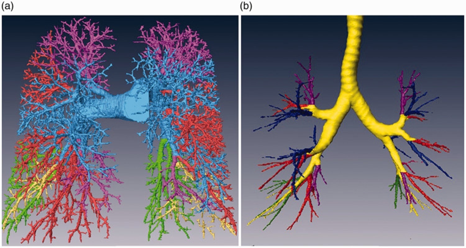
Branches of the (a) pulmonary artery and (b) pulmonary bronchi. Image Credit: J Int Med Res. 2021 May; 49(5): 03000605211014383.
The researchers discovered that around 90% of pulmonary segments could be easily identified via visual counting in the reconstructed 3D images from healthy subjects.
Here, each segmental artery closely accompanies the segmental bronchus, with apparent gaps between each of the adjacent segmental arteries and without branching of the segmental arteries into the adjacent pulmonary segments.
However, in 10% of segments, segmental arteries did enter the adjacent pulmonary segments. These would be difficult to assess using conventional methods.
However, the high-resolution CT images, together with the 3D reconstruction method, enabled the team to attain a clear visualization of the segmental arteries and segmental bronchi in any anatomical direction.
The researchers could measure the pulmonary arteries and the intersegmental vein surrounding each segment. Identifying these structures is extremely crucial to allowing these blood vessels to be ligated during surgery.
The inaccurate identification of overlapping pulmonary arteries could result in bleeding during surgery and surgical failure.
In patients with pulmonary masses, the team identified the site of the mass in any imaging direction, and the size of the pulmonary mass was quantified using Amira Software.
A marked improvement
The researchers promote this approach as it significantly improves existing methods and could be utilized before and during operations to deliver detailed anatomical understanding to surgeons.
“In short, our novel method provided the detailed anatomical structure of every segment before pulmonary segmentectomy. This is a great improvement that can be used to guide operations, even intraoperatively,”6 the authors write in the Journal of International Medical Research.
However, the authors also state that the semi-automated approach is highly time-consuming and requires up to two hours of radiologist time per 2-3 segments. The automated approaches for identifying pulmonary segments have not been adequately precise for clinical use.
A completely automated segmentation method that enables the demarcation of arteriopulmonary segments would be a much-welcomed development in the future.
Thermo Fisher technology
The researchers employed the semi-automated segmentation tools offered by the Thermo Fisher Amira Software for this study to conduct extraction of the structures of interest in all three anatomical planes.
The software facilitated the computation of the length, diameter, and angle of the pulmonary arteries, in addition to the quantification of the size of pulmonary masses in lung cancer patients.
This powerful software delivers 2D-5D image visualization, processing, and analysis from several modalities, such as optical and electron microscopy, MRI, and CT. As a flexible software solution, Amira Software provides a wealth of in-built tools together with the capability to customize its functions to specific needs.7
References and further reading
- American Cancer Society (ACS). Key statistics for lung cancer. Available at: https://www.cancer.org/cancer/lung-cancer/about/key-statistics.html. Accessed January 2023.
- Okada, M. et al. Correlation between computed tomographic findings, bronchioloalveolar carcinoma component, and biologic behavior of small-sized lung adenocarcinomas. J Thorac Cardiovasc Surg. 2001;71:3:956-960. Doi: 10.1016/j.jtcvs.2003.08.048
- Okada, M, et al. Is segmentectomy with lymph node assessment an alternative to lobectomy for non-small cell lung cancer of 2 cm or smaller? Ann Thorac Surg. 2020:143:115620. Doi: 10.1016/s0003-4975(00)02223-2
- Landreneau, RJ, et al. Recurrence and survival outcomes after anatomic segmentectomy versus lobectomy for clinical stage I non-small-cell lung cancer: a propensity-matched analysis. Journal of Clinical Oncology. 2014;32:23:2449-2455. Doi: 10.1200/JCO.2013.50.8762
- Fu H, et al. The arterial-ligation-alone method for identifying the intersegmental plane during thoracoscopic anatomic segmentectomy. J Thorac Dis. 2020;12:2343-2351. Doi: 10.21037/jtd.2020.03.83.
- Gao H, Liu C. Demarcation of arteriopulmonary segments: a novel and effective method for the identification of pulmonary segments. J Int Med Res. 2021;49:3000605211014383. Doi: 10.1177/03000605211014383.
- Amira software for Anatomical Biology Thermo Fisher Scientific - US. Available at: https://www.thermofisher.com/de/de/home/electron-microscopy/products/software-em-3d-vis/amira-software/pre-clinical-research.html
Familiarize yourself with Amira Software for bone and tissue image analysis
With its advanced image processing, segmentation, quantification, and reporting features powered by artificial intelligence (AI), Amira Software can provide you with the tools you need to take your discoveries to the next level.
In this webinar, you will discover how it was used to:
- Easily identify and analyze structural bone regions and precisely examine and measure specific bone structures to gain a deeper understanding of bone health, growth patterns, diseases, or responses to various treatments
- Perform advanced characterization of the growth plates in murine knee bones to obtain valuable insights into skeletal development, growth disorders, and potential treatments for related conditions
- Obtain quantitative information of bone tissue microstructure to understand bone biomechanics and the impact of different factors on bone structure and function
Watch on-demand Webinar
Sponsored Content Policy: News-Medical.net publishes articles and related content that may be derived from sources where we have existing commercial relationships, provided such content adds value to the core editorial ethos of News-Medical.Net which is to educate and inform site visitors interested in medical research, science, medical devices and treatments.