Peyer’s patches (PPs) are clusters of lymphatic tissue in the small intestine that act as our body’s first line of defense against ingested pathogens; they contain large amounts of white blood cells (T and B cells) to detect and process infectious agents, triggering an immune response.1
While these patches are known to form prenatally, how they continue to develop after birth is still being investigated. Understanding how PPs change in response to dietary, microbial, and environmental factors could help explain their maturation, particularly during weaning, and guide the development of therapeutics that support their function.
3D reconstruction and analysis of lymph follicles
Histological investigation of PPs using 2D cross-sections provides limited information because the lymph follicles (LFs) of the PPs are round. Cross-sections through circular structures look similar regardless of where they are taken, making it difficult to determine what depth (i.e., the center, margins, etc.) is being visualized. As a result, efforts have been made to analyze PPs in 3D in order to obtain a more holistic view of the lymphatic structures.
Serial 2D sectioning combined with 3D reconstruction is a practical approach that enables the visualization of volumetric data, and is used extensively across a wide variety of samples.
The success of these reconstructive methods depends heavily on the software used to align and superimpose the 2D data. A recent study, led by researchers at Tohoku University, Japan, utilized Thermo Scientific Amira Software in combination with microtome serial sectioning to image the Peyer’s patches of mice at different stages of maturity (between 2 and 10 weeks). They hoped to obtain a clearer picture of the postnatal development of the lymphatic tissue and deeper insights into its general organization and structure.
Novel insights into the maturation of post-natal Peyer’s patches
3D reconstruction in Amira Software revealed a number of novel insights that would have otherwise been unobtainable in 2D. The team found that the number of LFs remained largely unchanged over the 10 weeks; instead, the follicles increased in size, indicating a continuous recruitment of immune cells after birth.
After weaning, all LFs developed germinal centers (GCs) and the number remained constant consistent between 4 and 10 weeks. However the size of the GCs increased at 10 weeks compared to 4 weeks.
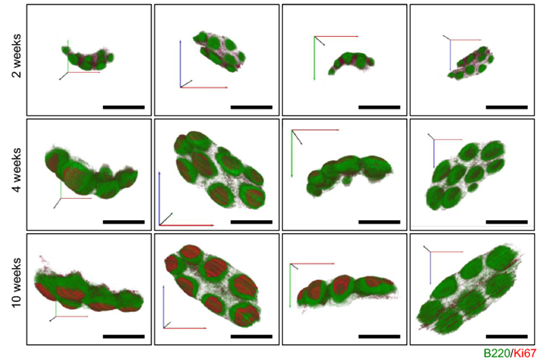
3D visualization of mouse Peyer’s patches 2-10 weeks after birth, shown from 4 different angles. Reconstruction was performed with Amira Software. Green: lymph follicles containing B220+ B cells. Red: Germinal centers including Ki67+ proliferating cells. Image Credit: Figure reproduced in part from Teshigahara et al. under CC BY 4.0.
Additionally, they found that the follicles had distinct interconnection points within the patches. Previously, LFs were thought to be largely independent, but these adhesion points appear to develop early on, and their presence did not change as the mice aged. These findings suggest that the follicles may leverage these adhesion points to facilitate the exchange of immune cells within the patches.
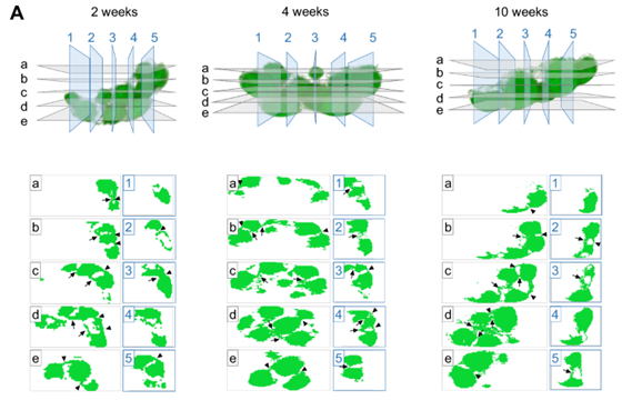
Analysis of junctions between lymph follicles in Peyer’s patches. Cross-sections taken from 2-10 weeks show that the number of junctions stays relatively static throughout the development of the patches. Image Credit: Figure reproduced in part from Teshigahara et al. under CC BY 4.0.
Finally, Teshigahara et al. were able to utilize biomarkers of cell proliferation to determine where new PP development was occurring. They found that GCs containing Ki-67+ antigen were not distributed uniformly within the patches, and instead were found to concentrate closer to the intestine’s muscle tissue, indicating patch growth in that direction. This could potentially be done to facilitate the activation and proliferation of immune cells from the patches.
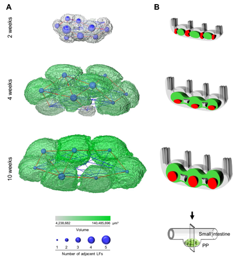
3D analysis of Peyer’s patches in Amira Software. A) Lymph follicle volume is shown in green, with junctions between LFs shown in blue. B) Germinal cell development (red) indicates that PPs develop preferentially near the muscle layer. Image Credit: Figure reproduced from Teshigahara et al. under CC BY 4.0.
Conclusions
Overall, 3D reconstruction in Amira Software enabled Teshigahara et al. to obtain a number of novel insights into the post-natal development of Peyer’s patches in the small intestine of mice.
The ability to align and correlate 2D serial sectioning data, along with segmentation of critical patch regions (i.e., luminal, middle, muscular, and interfollicular), makes Amira Software a powerful solution for this type of characterization.
More broadly, the insights provided by 3D reconstruction are revealing vital information on the composition, structure, and development of tissues. These unique observations greatly expand our understanding of biological functions, paving the way for the development and optimization of treatments and therapeutics targeting a wide variety of diseases and disorders.
Learn more about the 3D reconstruction and segmentation of tissues at thermofisher.com/amira
References
- Newberry, R.D. and Lorenz, R.G. (2005). Organizing a mucosal defense. Immunological Reviews, 206(1), pp.6–21. https://doi.org/10.1111/j.0105-2896.2005.00282.x.
- Anri Teshigahara, Banba, Y., et al. (2024). Formation of the junctions between lymph follicles in the Peyer’s patches even before postweaning activation. Scientific Reports, 14(1). https://doi.org/10.1038/s41598-024-65984-4.

Sponsored Content Policy: News-Medical.net publishes articles and related content that may be derived from sources where we have existing commercial relationships, provided such content adds value to the core editorial ethos of News-Medical.Net which is to educate and inform site visitors interested in medical research, science, medical devices and treatments.