Higher Resolution Microscopy Creates a Large Data Problem
Since their invention in the late sixteenth century, microscopes have been an invaluable tool to help life scientists study the complex structures and dynamic processes of living organisms.

Image Credit: Data courtesy of Dr. Srigokul Upadhyayula, Howard Hughes Medical Institute (HHMI) and Harvard Medical School
Microscopes evolved over time from basic magnification instruments to very advanced and highly selective optical microscopes and high-resolution electron microscopes. Other imaging techniques like computed tomography (CT), magnetic resonance imaging (MRI) and positron emission tomography (PET) became available in the second half of the 20th century.
A key criterion in biological imaging is the resolution limit; the ability of a microscope to differentiate between separate components. Light microscopy enables observation of live samples in real time but is limited by its inability to resolve subcellular structures. In contrast, EM offers much higher resolution but requires a vacuum to achieve this detail and so cannot be used to analyze live samples. Therefore, while each technique can be used alone, a combination of methods is often implemented to garner more detailed information. However, with more data collections techniques available, the size of data sets grow larger and larger. These data sets need to be processed and analyzed into understandable, quantitative forms for researchers to draw meaningful conclusions. Thus, extracting relevant information from large data sets from high resolution microscopy poses a large data challenge to scientists.
Microscopy Tools and Techniques are Continuously Advancing
Historically, microscopy has been performed on isolated cells adhered to glass and has enabled scientists to gather a wealth of information. However, as these samples are not in their native environment with this method, true physiological imaging is not possible. Furthermore, microscopy provides information of a snapshot in time, so analysis is moving towards five-dimensional (5D) imaging; dynamic 3D images (4D) that are acquired at multiple time steps. Therefore, much research and innovation have been invested to achieve this goal.
One such development was reported by researchers at the Howard Hughes Medical Institute, USA. In a 2018 paper published in Science, the Liu et al. group combined lattice light-sheet microscopy (LLSM), which offers non-invasive in vivo imaging at high spatiotemporal resolution, and adaptive optics (AO), which corrects for sample-induced aberrations commonly observed in multicellular specimens to improve resolution.
Higher Resolution Microscopy Generates Vast Amounts of Data
While these innovations in microscopy allow researchers to move closer to observing specimens in their fully native physiological state, it produces a new challenge; the size and volume of raw data generated for each analysis are dramatically increased.
To obtain as much meaningful insight as possible from these vast data sets, the information must be stored initially as raw files; deconvoluted, creating a second set of files; and then processed using imaging software, creating a third set of files. This processing can take many hours and often only produces a small amount of useable data, preventing meaningful real-time feedback on whether the samples are being optimally imaged. In fact, while demonstrating the combined AO-LLSM technique, researchers at the Howard Hughes Medical Institute, USA, generated 0.62 terabytes of raw data, only a minute fraction of which was presented.
This abundance of data can cause scientists’ workstations to struggle, so using imaging software specifically designed for high-resolution, multichannel, and time series data is essential to accurately produce usable results and draw conclusions from large data. Imaging software supports innovation, allowing laboratories to overcome limitations of copious amounts of information from data acquisition processes.
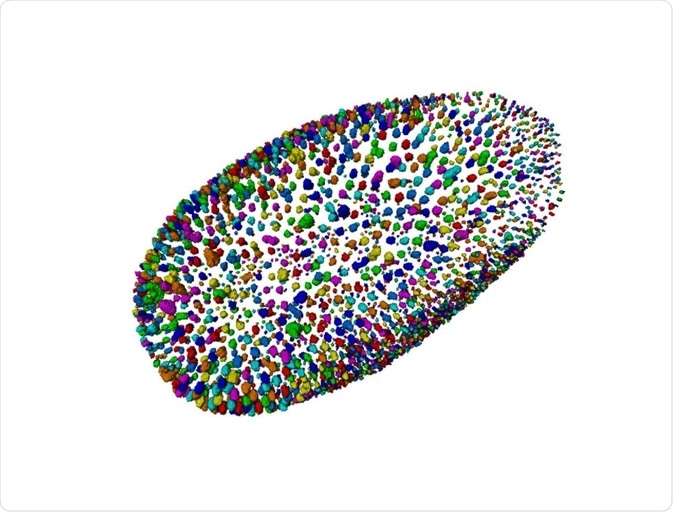
Image Credit: Data courtesy of Dr. A. Jain, Max Planck Institute of Molecular Cell Biology and Genetics, Dresden, Germany
Thermo Scientific’s Amira™ Software Extension – Xplore5D – Can Meet the Demands of Large Data Image Processing
To meet these needs, Thermo Scientific has just launched a new extension to their Amira Software – Xplore5D. Amira Software is a powerful platform for visualizing, manipulating, and processing imaging data. Amira Software has been supporting microscopy imaging analysis for decades. With the Xplore5D extension the handling of large, multichannel, and time series data for visualization, correlation and processing is possible.
Amira Software can process images at any scale. The Xplore5D extension can be used with many imaging modalities, including light sheet fluorescence microscopy, EM, CT, MRI, PET, and others, and is designed to be used for the entire workflow, from acquisition and segmentation to presentation of results. The cutting-edge, correlative analysis enables visualization of a range of acquired images across resolution and scale in one platform.
Xplore5D has a smart file format, allowing users to compress all imaging data without losing precious information collected during acquisition. This fast and flexible file conversion allows for immediate visual feedback so researchers can interact quickly and easily with the imaging data. The high-quality renderings enable users to build interactive 3D volume visualizations and animations to aid in analysis, communication, and collaboration.
Amira Software comes with artificial intelligence deep learning capabilities, which can improve image resolution, noise and offers automated segmentation. Furthermore, it enables complex workflows to be transferred into simple, step wise processing methods for future use or non-imaging specialists. With the Xplore5D extension, new multi-class or multi-label deep learning features have been added, including a training module and a prediction module.
These features enable researchers to easily work with large, multichannel and time series data sets from a wide range of high-resolution microscopy techniques on one platform to accelerate and streamline imaging analysis. The combination of high-resolution 5D microscopy with this powerful imaging software gives life scientists the tools to answer complex research questions across structural and cellular biology, tissue imaging, neuroscience, preclinical imaging, and bioengineering.
References