The brain is the most energy-demanding organ in the body, in part due to its complexity. Its components are varied and intricate: comprising different cell types, including neurons designed to transmit information, immune cells designed to protect brain functions, astrocytes designed to regulate the chemical environment, and glia designed to provide structural and functional support.
The overall functionality, health, and homeostasis of the brain are also impacted by non-cellular components, such as the (g)lymphatic system and cerebrospinal fluid (CSF). The brain has become a key area of scientific research because of its vital role in the body, among other things.1
Light-sheet microscopy is helping to overcome several challenges in brain imaging. It enables researchers to study a wide range of brain tissues and structures, including organoids, the eye, the spinal cord, and the peripheral nervous system.
Challenges in brain imaging
The brains intricate, complex nature makes it extremely difficult to study.
With countless cells working together across the organ, there is often a trade-off between achieving high-resolution images and being able to image the entire brain. It’s even harder to image the brain in its non-embryonic stages, particularly in humans and other vertebrates, because of the skull that surrounds it.
Developments in imaging are often a result of advancements in technology. For example, in medicine, functional magnetic resonance imaging (fMRI) is one of the gold standards for brain imaging in humans.2
Deep learning and artificial intelligence are having a massive effect on medical imaging, rapidly developing the field with their data acquisition and analysis capabilities.3
Changes in sample preparation techniques, like brain expansion, often come into play when working with biomedical and preclinical models. Issues with resolution can be overcome by physically expanding brain tissue, increasing the sample size instead of decreasing resolution.
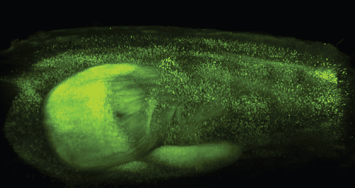
Figure 1. Neurons of a transgenic mouse expressing fluorescent protein YFP. The brain was cleared with Clarity. Image Credit: Dr. Zhang Dan, Tsinghua University, China.
Another challenge in brain imaging is the brain’s opacity in most organisms. This limits light penetration and makes it difficult to capture clear images beyond the surface.
Tissue clearing helps overcome the challenge of brain opacity by making tissues transparent, allowing light to penetrate deeper. This technique enables detailed imaging of internal brain structures, including complex neural networks.4
Neurobiology versus neurodevelopment
Two significant areas of interest in brain research include brain development and neurobiology.
Brain development research examines the processes of brain formation and its maturation throughout different stages of life, providing key insights into how brain disorders can stem from abnormalities during development.
Neurobiology research examines brain function, including decision-making, memory, and sensory perception. It also includes investigations into neuroplasticity, neurotransmitters, and neurochemistry. A thorough understanding of neurobiology is essential in diagnosing and treating a wide range of neurological conditions.
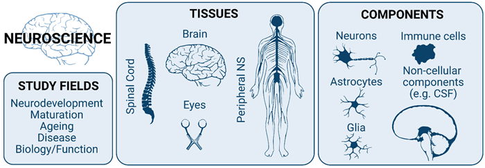
Figure 2. The term neuroscience encompasses different study fields, tissues, and nervous system components. Image Credit: Bruker Nano Surfaces and Metrology
Light-sheet fluorescence microscopy in brain imaging
Light-sheet fluorescence microscopy (LSFM) is an extremely effective tool for brain imaging, offering an array of practical benefits in neuroscientific research. Its key features include reduced phototoxicity, penetration depth, and the capacity to image rapid processes.5
LSFM's versatility becomes clear when considering its applications for the live imaging of dynamic processes, such as long-term developmental changes in the brain or calcium dynamics. It is also ideally suited to the imaging of cleared and expanded tissues, offering cellular-level resolution of entire brains.6
Hindbrain morphogenesis
A study into the role of Notch-3 in the neurogenic fate of hindbrain boundaries saw intricate time-lapse imaging and data analysis used to highlight specific boundary cells during hindbrain morphogenesis depending on Notch-3 signaling.7
This research leveraged Bruker’s Multi-View Selective-Plane Illumination Microscope (MuVi SPIM) for cell lineage analysis.
Imaging beyond the brain: The eye
Eye research is central to understanding and preventing loss of vision, but it is also regularly used as a proxy for understanding the brain. This is because, despite the eye being peripheral, it is a much more accessible part of the brain.
The eyes and brain share many similarities regarding cell types, functionality, and cell-to-cell interactions. The eye is an invaluable model for fundamental biological processes due to its structural and functional resemblance, with eye research not only critical in itself but also as a bridge to wider neurology research.8
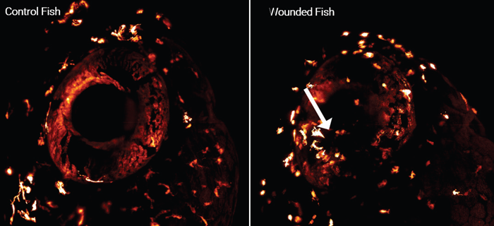
Figure 3. Microglia and macrophage dynamics after wounding with photomanipulation. Image Credit: Dr Gordon Wang, Stanford University, USA.
Subcellular mouse retina dynamics
Mouse retinas were imaged in 3D using a MuVi SPIM to study subcellular Golgi apparatuses and tuft morphology.9
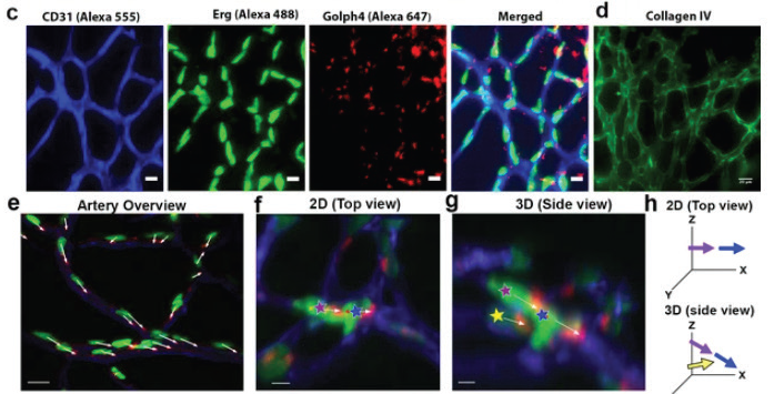
Figure 4. Light-sheet fluorescence microscopy was used to study subcellular components and cellular polarization. (c) Retina stained with antibodies for blood vessels using anti-CD31(Alexa 555), vascular nuclei Anti-Erg (alexa488), and Golgi apparatus using anti-Golgi (Golph4, Alexa 647). (d) Collagen IV stained retina. (e) Image showing aligned cell polarization. Figure 1 - figure supplement 1 reproduced under CC BY 4.0 DEED license.9 Image Credit: Bruker Nano Surfaces and Metrology
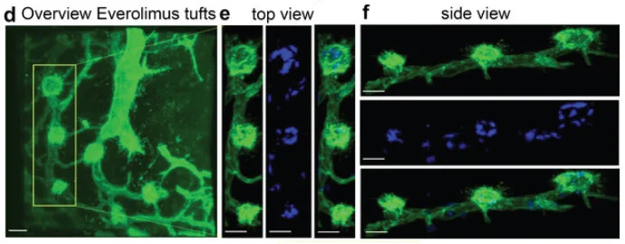
Figure 5. Light-sheet fluorescence microscopy shows 3D tuft morphology. mTOR inhibition with Everolimus leads to highly active filopodia and cup morphology in vascular tufts. Figure 6 - figure supplement 3 reproduced under CC BY 4.0 DEED license.9 Image Credit: Bruker Nano Surfaces and Metrology
Imaging beyond the brain: The spinal cord
The spinal cord plays an essential role in carrying signals between the brain and the rest of the body, influencing essential functions such as sensation, movement, and organ control.
Developments in spinal cord research could potentially unlock new treatments and therapies for patients with spinal cord injuries, neurological disorders, and paralysis.10
Imaging the peripheral nervous system
Scientists can achieve unprecedented access to the intricate details of peripheral nerves by studying the peripheral nervous system (PNS), which helps map out the structural intricacies of nerve fibers and visualize the dynamic processes that are essential for better understanding neural function.
Biomedical imaging helps reveal the connections between the peripheral nervous system (PNS) and the brain. It allows researchers to trace neural pathways and explore how information is processed and transmitted throughout the nervous system.
Studying the PNS is fundamental to the diagnosis and monitoring of neurological disorders affecting both peripheral nerves and the brain.11 This integrated approach provides vital insights with the potential to advance medical diagnostics, treatment strategies, and wider neurological research.
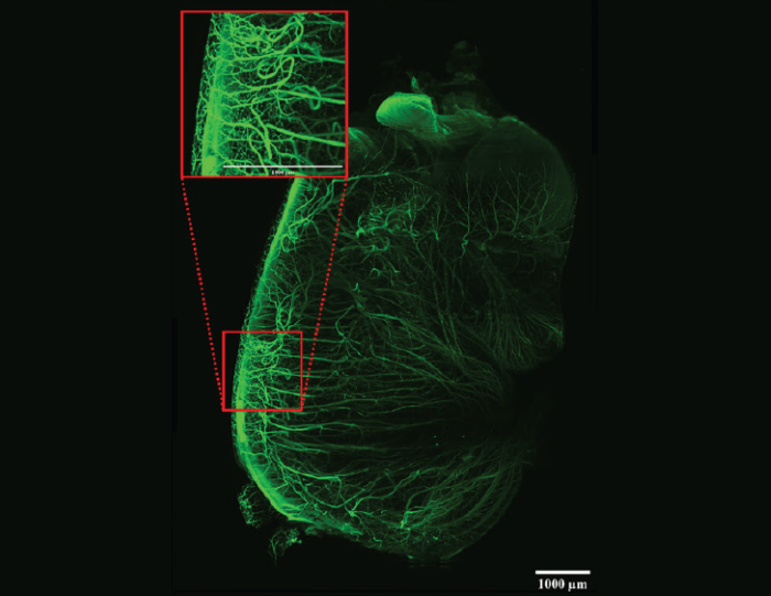
Figure 6. Developing nerves in a whole mouse embryo. The sample was cleared with DBE. Tiled image (3 x 4) acquisition. Scalebars: 1 mm. Imaged on the LCS SPIM. Sample courtesy of James Muller, MSKCC, New York, USA. Image Credit: Bruker Nano Surfaces and Metrology
Cell culture and organoids
Three-dimensional cell cultures are fragile systems that require low phototoxicity during imaging and optimized experimental protocols to preserve delicate samples.
Light-sheet imaging offers a solution to these challenges, allowing researchers to study cell culture systems in 3D, including time-lapse imaging.
Astrocyte incorporation into neuronal organoids
One research project sought to establish human cell-based neural organoids, demonstrating that the addition of astrocytes resulted in the creation of a micro-physiological system.12 The researchers used the MuVi SPIM microscope to image organoids during this study.
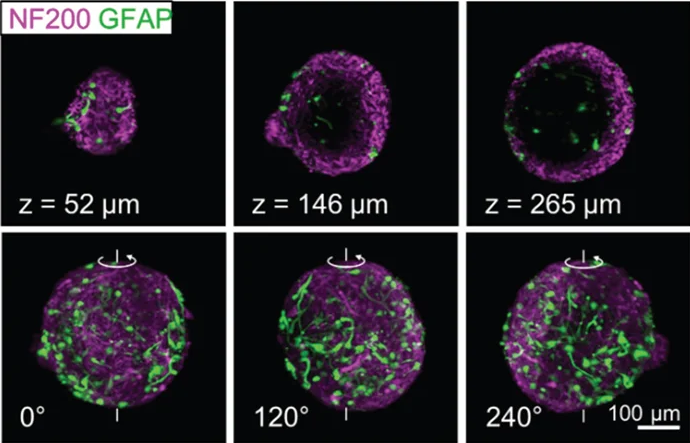
Figure 7. Co-cultured organoids with neuronal cells (magenta) and astrocytes (green). The top row shows slices, and the bottom row shows 3D reconstructions and stills of rotations of organoids. Figure 4 - altered from red-green to magenta-green to be colorblind-friendly. Adapted and reproduced under Creative Commons Attribution 4.0 International License (shared with author permission).12 Image Credit: Bruker Nano Surfaces and Metrology
Conclusion
As the body’s central control hub, the brain plays a vital role in overall health, making it vital to understand how it functions. Despite its complexity, innovative tools and techniques are steadily deepening our knowledge of brain structure and activity.
This cutting-edge research opens the door to major advances in brain development and neurobiology, with the potential to improve diagnosis, treatment, and quality of life for those affected by neurological disorders.
The Best Light-Sheet Microscopes for Different Neuroscience Applications. Source: Bruker Nano Surfaces and Metrology
| Use Case |
Scientific Requirements |
Bruker Microscope |
Large
Cleared
Samples |
- Brain mapping or connectivity analysis
- Whole-body nerve assessment
- Whole organ studies
|
LCS SPIM
Large, cleared sample light-sheet fluorescence microscope |
Fragile
Samples |
- Organoid differentiation
- Organ-explant studies
- Fast imaging of developmental processes
- optional photomanipulation module
|
TruLive 3D Imager
Dual-sided illumination light-sheet fluorescence microscope |
Live, Fixed, or
Cleared Samples |
- Live, long-term or cleared sample imaging
- Multi-view imaging without rotation
- Optional photomanipulation module
|
MuVi SPIM
Multiview imaging for live and cleared samples |
Specialized
High-Resolution
Imaging |
- High-resolution imaging of live or fixed samples
- Variable beam patterns
- Allows for specialized applications, such as SIM, FLIM, or FCS
- Optional photomanipulation module
|
InVi SPIM Lattice Pro
Inverted view light-sheet fluorescence microscope with advanced illumination |
References and further reading
- M. Bear, B. Connors, and M. A. Paradiso, Neuroscience: Exploring the Brain, Enhanced Edition: Exploring the Brain, Enhanced Edition. Jones & Bartlett Learning, 2020.
- Poldrack, R.A. and Farah, M.J. (2015). Progress and challenges in probing the human brain. Nature, (online) 526(7573), pp.371–379. https://doi.org/10.1038/nature15692.
- Suri, J.S. (2019). State-of-the-art review on deep learning in medical imaging. Frontiers in Bioscience, 24(3), pp.392–426. https://doi.org/10.2741/4725.
- Murakami, T.C., et al. (2018). A three-dimensional single-cell-resolution whole-brain atlas using CUBIC-X expansion microscopy and tissue clearing. Nature Neuroscience, 21(4), pp.625–637. https://doi.org/10.1038/s41593-018-0109-1.
- Huisken, J. (2004). Optical Sectioning Deep Inside Live Embryos by Selective Plane Illumination Microscopy. Science, 305(5686), pp.1007–1009. https://doi.org/10.1126/science.1100035.
- Ahrens, M.B., et al. (2013). Whole-brain functional imaging at cellular resolution using light-sheet microscopy. Nature Methods, (online) 10(5), pp.413–420. https://doi.org/10.1038/nmeth.2434.
- Hevia, C.F., et al. (2022). The neurogenic fate of the hindbrain boundaries relies on Notch3-dependent asymmetric cell divisions. Cell Reports, 39(10), pp.110915–110915. https://doi.org/10.1016/j.celrep.2022.110915.
- London, A., Benhar, I. and Schwartz, M. (2012). The retina as a window to the brain - from eye research to CNS disorders. Nature Reviews Neurology, 9(1), pp.44–53. https://doi.org/10.1038/nrneurol.2012.227.
- Prahst, C., et al. Mouse retinal cell behaviour in space and time using light sheet fluorescence microscopy. eLife, (online) 9, p.e49779. https://doi.org/10.7554/eLife.49779.
- Lewis, K.E. and Eisen, J.S. (2003). From cells to circuits: development of the zebrafish spinal cord. Progress in Neurobiology, 69(6), pp.419–449. https://doi.org/10.1016/s0301-0082(03)00052-2.
- J. Hubbard, The Peripheral Nervous System. Springer Science & Business Media, 2012.
- Brüll, M. (2020). Incorporation of stem cell-derived astrocytes into neuronal organoids to allow neuro-glial interactions in toxicological studies. ALTEX. https://doi.org/10.14573/altex.1911111.
Acknowledgments
Produced from materials originally authored by Dr Elisabeth Kugler from Bruker.
About Bruker Nano Surfaces and Metrology

Bruker’s suite of fluorescence microscopy systems provides a full range of solutions for life science researchers. Their multiphoton imaging systems provide the imaging depth, speed and resolution required for intravital imaging applications, and their confocal systems enable cell biologists to study function and structure using live-cell imaging at speeds and durations previously not possible. Bruker’s super-resolution microscopes are setting new standards with quantitative single molecule localization that allows for the direct investigation of the molecular positions and distribution of proteins within the cellular environment. And their Luxendo light-sheet microscopes, are revolutionizing long-term studies in developmental biology and investigation of dynamic processes in cell culture and small animal models.
Sponsored Content Policy: News-Medical.net publishes articles and related content that may be derived from sources where we have existing commercial relationships, provided such content adds value to the core editorial ethos of News-Medical.Net which is to educate and inform site visitors interested in medical research, science, medical devices and treatments.