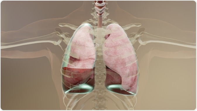
Empyema is considered to be a sub-classification of parapneumonic pleural effusion. A parapneumonic effusion describes the build-up of activated pleural fluid which is associated with a lung infection, predominantly pneumonia.

Image Credit: ALIOUI MA/Shutterstock.com
Determining the underlying cause is facilitated by several processes which include thoracentesis and pleural fluid analysis. The plural fluid, And chloral infusions at large, may be classified as transudate or exudate, depending on etiology. Pleural effusion is an umbrella term used to describe any abnormal accumulation of fluid in the pleural cavity.
An empyema can resemble a pleural effusion and can mimic a peripheral pulmonary abscess. As such, diagnosis is important in distinguishing the features that may help distinguish empyema from other forms of pleural effusion. Empyema is characterized by a frank post in the pleural space, presence of bacterial infection of the pleural fluid as ascertained by gram stain or positive culture.
This form of pleural effusion is common in patients who developed pneumonia. Between 40 and 60% of patients with bacterial pneumonia are unlikely to develop a form of pleural effusion of varying severity.
Initial evaluation of pleural effusion including empyema
The history and physical examination of the patient are critical in guiding the evaluation of pleural effusion. These include:
- A history of abdominal surgical procedures in which post-operative pleural effusion, subphrenic Abscess, and pulmonary embolism may be the cause of empyema
- A history of alcohol abuse or pancreatic disease which may cause a pancreatic effusion artificial pneumothorax therapy in which pyothorax-associated lymphoma, trapped long and tuberculosis empyema may result
- history of cancer in which malignancy can cause empyema
- cardiac surgery or myocardial injury in which any form of pleural effusion, including empyema, occurs secondary to coronary artery bypass graft surgery or Dressler’s syndrome
- chronic hemodialysis which may cause heart failure and uremic pleuritis
- cirrhosis which can cause hepatic hydrothorax, and spontaneous bacterial empyema
- Childbirth, which can cause postpartum pleural effusion, including hepatic hydrothorax, and spontaneous bacterial empyema
Signs and symptoms of an effusion vary depending on the underlying disease, but shortness of breath (dyspnoea), cough, and pleuritic chest pain are the most common signs. Consequently, an examination of the chest of a patient with suspected pleural effusion, including empyema is essential for detecting hallmarks of the condition.
These include dullness to percussion, decreased breathing sounds, no voice transmission, and an absence of tactile fremitus. Tactile fremitus refers to the vibration of the chest wall that results from sound vibrations created by speech or other vocal sounds. Other signs include:
- Ascites
- Pericardial friction rub resulting in pericarditis (swelling and irritation of the thin, saclike tissue surrounding the heart (pericardium))
- Peripheral edema, elevated jugular venous pressure which can manifest as heart failure and constrictive pericarditis
- Swelling of the lower extremities
- Yellowing nails and lymphoedema
Symptoms of empyema include fever, hemoptysis (coughing up blood), and weight loss.
Confirming suspected empyema
Due to a lack of specific diagnostic criteria due to the variability in clinical presentation, several tests are needed to establish a diagnosis of empyema. The first test implemented is a chest x-ray. This form of testing is widely available and simple, although it lacks sensitivity.
A threshold level of fluid must be detected; usually 75ml in the lateral view and 175ml in the anterior view, Moreover, some features of the pleural effusion may be blunted due to costodiaphragmatic (involving the ribs and diaphragm) angles, and lungs radiolucent fluid present in the lungs (transparent to X-rays).
Ultrasound
If an effusion is suspected with x-ray evaluation, an ultrasound is typically conducted. As with x-ray, this technique is widely available and easy to administer; it offers relatively greater sensitivity in the detection of pleural effusion as compared to x-rays, allowing differentiation between parenchyma and pleural fluid. It may also offer a therapeutic benefit, providing a useful guide to chest tube placement during thoracentesis.
Computed tomography (CT) scan
CT scan of the chest may be an alternative option after a chest x-ray or ultrasound. A CT scan is performed with intravenous (IV) contrast to enhance the pleura. This helps differentiate lung abscess from empyema and transudate from exudate.
Transudate is extravascular fluid with low protein content, where is exudate is comparatively higher in protein content. A transudative pleural effusion occurs due to increased hydrostatic pressure or low plasma oncotic pressure whereas exudative pleural effusion occurs due to inflammation and increased capillary permeability.
Visualization of the pleural surfaces is possible, and this helps identify pleural fluid locations. The presence of empyema is suggested by a split pleura sign in which there is a thickening of the two forms of pleura the visceral and parietal pleura, with significant separation of both pleural services.
CT is more accurate to separate empyema from underlying compressed lung than a plain chest radiograph, and CT thorax is more accurate for diagnosis and is better at characterizing the size and location of a pleural effusion. A CT is useful as a guidance tool to locate the skin entry sites for thoracentesis or drainage catheter placement when ultrasound is limited in the case of cases where there are adjacent bony structures, in a large patient, or when there is air in the lung parenchyma.
Thoracentesis
To guide the management of parapneumonic pleural effusion, diagnostic thoracentesis is performed. Thoracentesis is a valuable procedure that enables a fluid sample to be obtained for differentiating transudate from the exudate and allowing the removal of the fluid in a patient with a large volume of effusion for symptomatic relief.
The most common diagnostic characteristic seen in a thoracentesis is fluid in the pleural space which occupies more than 10mm in thickness on lateral decubitus radiograph with unknown etiology. A lateral decubitus abdominal radiograph is performed in a patient when the patient is lying on either the left (left lateral decubitus) or right (right lateral decubitus) side.
The obtained fluid is then sent for analysis and culture. The pleural fluid is subject to microbiological analysis using gram staining with cultures, pleural fluid total and differential cell count, biochemical analysis which reveals the total protein, lactate dehydrogenase, glucose, and pH; and pleural fluid biomarkers which include biomarkers of infection for example C reactive protein which I used to distinguish complicated parapneumonic pleural effusions from uncomplicated parapneumonic pleural effusions.
References:
Further Reading
Last Updated: Mar 14, 2023