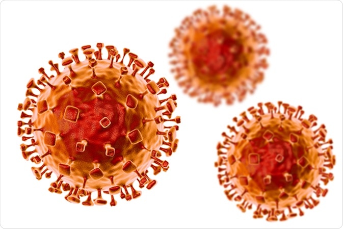
Infections with Nipah virus can affect both humans and animal reservoirs. The presence of Nipah virus is confirmed using several levels of testing, which primarily includes viral isolation, as well as serology and nucleic acid amplification, the latter of which is also known as molecular diagnostics.

Image Credit: Kateryna Kon / Shutterstock
Isolation
Biosafety level (BSL)-4 laboratories are required for the isolation and propagation of the Nipah virus due to its intense deadliness. Primary isolation may be carried out using suspected samples for confirmation of infection in BSL-3 labs if stringent precautions are complied with to ensure the safety of the operators.
However, if the specimen is positive for viral DNA by immunofluorescence testing of infected cells fixed with acetone, the culture fluid must be transferred immediately to a BSL-4 laboratory.
Several high-security laboratories fulfilling BSL-4 criteria have emerged to deal with Nipah outbreaks in developing countries in view of its potential to become a fatal outbreak. These include:
- The OIE Reference Laboratory for Henipaviruses at Geelong for animal research in the Asia–Pacific region
- National Institute of Virology, ICMR, at Pune (India) and the High-Security Animal Disease Laboratory, Bhopal (India) for human and animal virus isolation facilities
- ICDDRB and IEDCR in collaboration with Centers for Disease Control and Prevention (CDC), USA
Serology
Several methods are in use for serologic diagnosis.
Viral antigen-capture enzyme-linked immunosorbent assay (ELISA) can process samples at high throughput while being cost-effective; therefore, this diagnostic technique is often considered highly useful for screening samples. On the other hand, monoclonal antibody-based antigen capture ELISA not only enables viral detection but may also be capable of distinguishing Nipah from Hendravirus, which is another highly pathogenic virus in the same family.
These methods are based upon the use of cell lysate antigens that are obtained from testing centers and can be performed only in BSL-4 laboratories. To get around this issue, alternative antigens have been developed in the form of recombinant proteins, such as the recombinant N protein ELISA.
In addition, there is a novel antigen-capture sandwich ELISA technique based on rabbit polyclonal antibody to NiV-G protein DNA vaccine, which is likely to help in the rapid diagnosis of new NiV that cannot be detected by conventional polymerase chain reaction (PCR) methods. Moreover, a novel serum neutralization technique is based on the use of pseudotype particles.
Methods of nucleic acid amplification
Infection with Nipah virus can also be detected using molecular diagnostic tests such as real-time or otherwise reverse transcriptase PCR (RT-PCR) and duplex nested RT-PCR. The results of these tests are confirmed by sequencing the products of DNA amplification.
Collection of samples, multiple testing, and antibody patterns
A patient with a clinical history that is suspicious of Nipah infection should be tested with multiple tests. During the early phase, virus isolation and real-time RT-PCR tests are carried out on swabs taken from the throat and nose, cerebrospinal fluid, urine, and blood.
In the later stages, immunoglobulin M (IgM) and IgG detection using serological tests, such as an ELISA, are done. If the patient dies before these tests are performed, samples of tissues obtained during the autopsy may need to be acquired for immunohistochemical testing to confirm the presence of the virus.
When serology tests are used for the diagnosis of Nipah infection, IgM antibodies are detectable during the first five days of symptomatic illness in approximately 66% of patients. In patients who survived, by 2 weeks IgM and IgG were universally positive.
By 2-3 months of onset, IgM levels started to fall and became negative in all survivors by 2 years, while IgG positivity remained unchanged in all patients at 2 years. Thus, a patient who is initially NiV negative by antibody testing may develop detectable levels of IgM antibodies by 2 weeks or more after the onset of illness.
Treatment
Being a novel virus with frequent mutations, no specific treatment is available at the moment for Nipah virus infection. Supportive care is mandatory to optimize survival.
Person-to-person transmission is documented in recent outbreaks, which mandates the use of standard precautions and barrier nursing methods when treating or diagnosing a suspected or confirmed case of Nipah infection. This is important, as almost half of Nipah virus patients in one outbreak were found to have contracted the virus nosocomially.
Ribavirin has been the only antiviral drug to date to show effectiveness against the Nipah virus; however, more studies are required to confirm this finding that has been largely based on in vitro data and few human studies.
Another drug that is being tested for the treatment of Nipah virus is a purine nucleoside analog that has already been approved in Japan for the treatment of new influenza virus strains. To date, this drug has been shown to be active against Hendra and Nipah viruses in vitro.
Another method being investigated is passive immunization using a human monoclonal antibody raised against the G-glycoprotein of the Nipah virus, which has shown significant benefit in studies.
References
Further Reading
Last Updated: Apr 13, 2021