This article is based on a poster originally authored by Bronwyn K Irving, Valeria Chichagova, and Carolina Gándara.
Photoreceptor outer segments (POS) are essential for vision, and their dysfunction is associated with retinal degeneration and subsequent vision loss.
Retinal organoids (ROs) have emerged as a valuable resource for investigating the mechanisms underlying retinal diseases and for drug discovery, due to their capacity to replicate certain features of the human retina.
A comprehensive understanding of the dynamics of POS development and the consistency across multiple batches of ROs is vital for their broader application and routine implementation.
The study presented here evaluates the dynamics, efficiency, and batch-to-batch reproducibility of POS development across three large-scale batches of wild-type human-induced pluripotent stem cell (iPSC)-derived retinal organoids.
Methods
Three independent batches of wild-type ROs were assessed for POS development.
Batches of 100 ROs were monitored over the differentiation timeline at days 120, 150, 180, 210, 240, and 270.
Image Credit: Newcells Biotech
Results
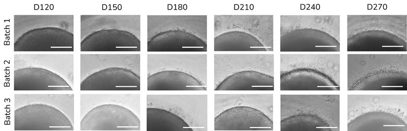
Figure 1. Over maturing timepoints POS increase in number, density and length. (A) Representative brightfield images of RO POS across 3 biological repeats. Scale bar 150 μm. Image Credit: Newcells Biotech
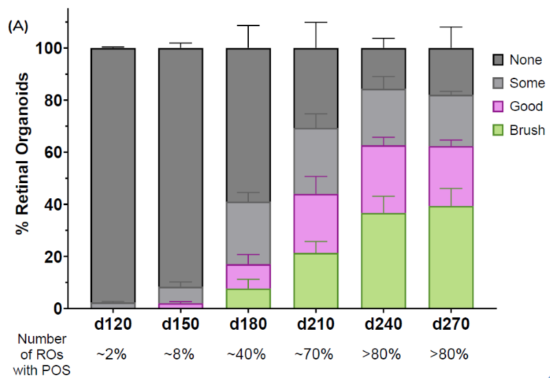
Figure 2. POS brush borders start appearing at ~Day 180, increase over time and plateau at Day 240. (A) 100 ROs scored in 4 categories for presence of POS across 3 batches with up to ±10% batch-to-batch variability (mean ± SEM). Percentages (%) indicate total number of ROs with presence of POS. Image Credit: Newcells Biotech

Figure 3. POS increase in presence and length over maturing time points at the protein level. (A) Expression of cone and rod cells, marked by OPNMLW and RHO respectively, increase over time, with the outer segment portion above the outer nuclear layer (marked by Hoechst). (B) Rod cell outer segments, marked by RHO, increase in length cross the outer limiting membrane, marked by ZO1. (C) Inner segments, marked by TOM20, protrude above the outer nuclear layer over time with an increase in photoreceptor outer segments marked by ROM1. (D) Connecting cilia, marked by ARL13B, increase in number and localize with the protruding TOM20 marked inner segments. Image Credit: Newcells Biotech
Conclusions
This study established the timeline for the development of POS in wild-type iPSC-derived ROs, as measured by bright field appearance and protein expression profiles across multiple large-scale batches.
The observations indicate that these large-scale batches of ROs consistently develop POS by day 240, making these more mature ROs suitable models for pre-clinical studies related to disease modeling, drug discovery, and gene therapy, where POS formation is pertinent.
Acknowledgments
Produced from material originally authored by Bronwyn K Irving, Valeria Chichagova, and Carolina Gándara from Newcells Biotech.
About Newcells Biotech
Newcells Biotech develops in vitro cell-based assays for drug and chemical discovery and development.
Using our expertise in induced pluripotent stem cells (iPSCs), cellular physiology, and organoid technology, we build models that incorporate the “best biology” for predicting in vivo behavior of new drugs.
Our experts have developed and launched assays to measure transporter function, safety, and efficacy in a range of cell and tissue types, including kidney, retina and lungs.
We have the capability to develop and implement protocols to measure cilia beat frequency and toxicity on small airway epithelial cells model, retinal toxicity and disease modelling on retinal organoids and retina epithelium, as well as drug transport in the kidney, DDI and nephrotoxicity across human and a range of preclinical species.
Sponsored Content Policy: News-Medical.net publishes articles and related content that may be derived from sources where we have existing commercial relationships, provided such content adds value to the core editorial ethos of News-Medical.Net which is to educate and inform site visitors interested in medical research, science, medical devices and treatments.
Last Updated: May 13, 2025
 Newcells Biotech raises additional funding from investors