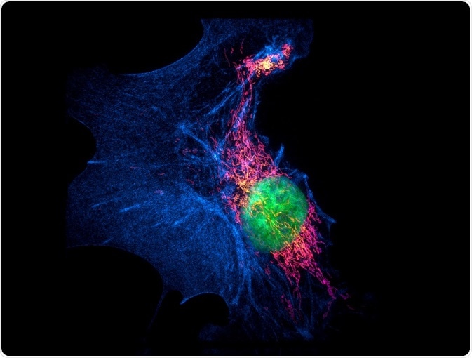
Super-resolution microscopy refers to a collection of methods used to increase the resolution of light microscopy, whereas confocal microscopy uses a laser beam to increase the signal intensity from the sample. These two types of microscopy have wildly differing approaches and applications, which confer different advantages and disadvantages.
Skip to:
 Micha Weber | Shutterstock
The Basics of Confocal and Super-resolution Microscopy
Super-resolution microscopy can come in different forms, but structured illumination microscopy (SIM) is one of the more common approaches. In this technique, grid projections are used to increase the resolution limit that is set by diffraction. The grid projections are cast on the sample at different angles and orientations. The subsequent fluorescent signal that is emitted from the sample is detected and reconstructed in the Fourier domain, where the final image is produced from the reconstruction.
Another common super-resolution microscopy technique is stimulated emission depletion microscopy (STED). The development of this technique built off the manipulation of fluorophores, rather than directing and focusing them. The STED technique allows for fluorophores outside of the imaging center to be switched off, thereby removing background noise or interfering signals.
The central idea of a confocal microscope is that the light is focused to increase the signal intensity from a sample. In many other types of microscopy, such as fluorescence microscopy, the excitation light targets the entire sample, leading to full fluorescence. This can be true for confocal microscopy as well, where more than the focal point is illuminated by a laser. However, confocal microscopes use a screen with a pinhole to target the focal point and only receive fluorescence from that part of the sample. This filters out other areas.
The word ‘confocal’ comes from this principle, where the pinhole is conjugate (the pinhole and the sample plane are conjugate planes) to the lens’ focal point. Once light has passed through the pinhole, it is measured by the detector. The detectors can come in different forms, but are generally photon detectors that convert the photons into electrical signals. These electrical signals can then be amplified, leading to higher contrast than many other microscopy methods.
Advantages of Super-resolution Over Confocal Microscopy
Differences in the applications of confocal and super-resolution microscopy translate to advantages in different areas. Confocal microscopes work well for imaging tissue and cell dynamics, but do fall short on the subcellular level. Super-resolution microscopy achieves better resolution at greater depths by controlling the modulation of the excitation light as opposed to light excitation in a uniform pattern.
Super-resolution microscopy is a relatively young method compared to confocal microscopy, and therefore developments are ongoing to improve standards. While confocal microscopy has been praised for its high contrast, recent methods show super-resolution microscopy, via the SIM technique, is capable of catching reflectance imaging with more than double the resolution of confocal microscopy. This increased resolving capability was applied to imaging nanoparticle uptake and localization inside cells. Such developments aid in super-resolution microscopy becoming a validated alternative to confocal microscopy for imaging subcellular activity.
.jpg) Vshivkova | Shutterstock
Vshivkova | Shutterstock
Advantages of Confocal Over Super-resolution Microscopy
Sample bleaching is a common issue for many microscopy techniques, super-resolution microscopy included. While confocal microscopy is often praised for its fast acquisition times of big sample areas, super-resolution microscopy is not as efficient. The extended measurement times associated with this method increase sample bleaching more than in some other microscopy techniques. This also impacts its feasibility in imaging live cells, though this can be bypassed by better technology that reduces the time needed to acquire image data.
Sample preparation can be more difficult in super-resolution microscopy than in confocal microscopy, due to deterioration of the illumination pattern in thicker samples. In most cases, samples need to be 20 microns or less to avoid deterioration when light passes through the sample. A further benefit of confocal microscopy is that live cells can be imaged, and cannot in super-resolution microscopy.
In experiments directly comparing confocal microscopy and super-resolution microscopy, confocal microscopy retains the edge when it comes to acquiring vast amounts of 3-dimensional data and with better detection sensitivity than super-resolution microscopy.
Sources
- Vangindertael J., et al. (2018). An introduction to optical super-resolution microscopy for the adventurous biologist. Methods and Applications in Fluorescence. http://doi.org/10.1088/2050-6120/aaae0c
- Guggenheim E.J., et al. (2016). Comparison of confocal and super-resolution reflectance imaging of metal oxide nanoparticles. PLoS ONE. https://doi.org/10.1371/journal.pone.0159980
- Weeks E.R. (2007). How does a confocal microscope work? http://www.physics.emory.edu/faculty/weeks//confocal/
Last Updated: Oct 9, 2019