By Jeyashree Sundaram, MBA
Fluorophore is the specific region or structural domain of a protein that exhibits fluorescence. In any fluorescence spectroscopy, fluorophores are the most significant part to image the protein–protein interactions.
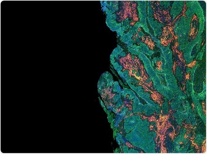
The edge of a tumour where the levels of different proteins has been probed for using antibodies labelled with fluorophores. Credit: Carl Dupont/Shutterstock.com
Various fluorophore proteins are available to image several biological processes, e.g., proteins sensitive to pH are used to assess pH, while fluorophore proteins with lengthy excitation and emission wavelengths can only be used in tissues, which spontaneously exhibit fluorescence at shorter excitation wavelengths. The technique involving interaction between the two fluorophores is used to understand the biological systems and it is termed as fluorescence resonance energy transfer (FRET).
Types of Fluorophores
Fluorophores are classified into two main groups: intrinsic and extrinsic. Fluorophores that are naturally obtained are termed as intrinsic, e.g., aromatic amino acids, derivatives of pyridoxal, flavins, NADH, and chlorophyll.
Fluorescence can also be produced by supplementing fluorophores to the sample that does not contain any fluorescent property or by altering the spectral properties of the sample. Such fluorophores that are used for artificially ingesting fluorescence are called as extrinsic fluorophores such as rhodamine, fluorescein, dansyl, and various other substances.
Fluorophore conditions in FRET analysis
FRET is a distance-dependent interaction between two fluorophores. In FRET, a light source excites a donor fluorophore and transfers the energy to an acceptor fluorophore without emitting light. The acceptor absorbs the energy and subsequently emits the visible light; using a specialized reader the loss and gain of fluorescence in donor and acceptor can be determined accurately. To achieve this, FRET has to meet several conditions.
Proximity
For efficient FRET processing, the donor and acceptor fluorophores must be kept close to one another.
Spectral overlap
The emitted energy of donor fluorophore must overlap the absorbed spectrum of the acceptor. The higher the intensity of spectral overlap the greater the possibility of FRET to occur.
Dipole orientation
Through intermolecular dipole–dipole coupling, energy shift takes place from the donor to acceptor fluorophore. The orientations of the dipole between the two fluorophores are commonly assumed to be random because of the fast molecular rotations.
FRET from several pathways in fluorophore-labeled DNA
DNA scaffolds give a beneficial approach for establishing fluorophores into complex antennae that harvest light. Though the size and DNA-fluorophore network’s complexity increases, it may be very challenging to understand the properties of energy transfer due to significant numbers of dipolar interactions among fluorochromes.
Using the combination of steady-state and time-resolved spectroscopy, FRET properties are illustrated and deciphered using Förster theory. Efficiency of FRET increases by multiple FRET pathways as the homo-FRET found between the donors permits access to parallel pathways to the acceptor, and thus, counteracts the low FRET efficiency.
In general, multiple pathways are required to design artificial devices to harvest light for compensating nonhomogeneous and nonideal group of effects that lower the FRET efficiency.
FRET analysis by quantum-dot fluorophores
FRET is a highly powerful photophysical tool that transfers nonradiative excitation energy to acceptor fluorophore at the ground state. It is highly sensitive to nanoscale changes in the distance between the donor and acceptor and their corresponding dipole orientations.
It has acquired a broad range of applications in protein conformation studies, analytical chemistry, and biological assays. Luminescent nanocrystals, called quantum dots, are inorganic fluorophores with distinct spectroscopic and photosensitive properties that could promote FRET as an analytical tool because of its wider excitation spectrum and symmetric and tunable fine photoemission. Utilizing FRET with luminescent quantum dots, studies focus on dealing with development of targeted applications in various studies.
Fluorophores used for FRET
CFP-YFP FRET pairs
Cyan–yellow fluorophore protein (CFP–YFP) pairs are the most common fluorophore protein FRET pairs. One benefit of cyan donors, such as mTurquoise2, mCerulean3, and Aquamarine, is that a few genetically engineered CFPs have high significant yield. They also possess greater fluorescence lifetime. The frequently used YFPs, such as mVenus, mCitrine, YPet, and sEYFP, are less responsive to chloride and pH, and highly stable to light exposure.
Conversely, CFP–YFP pairs experience numerous problems related to FRET, for instance fast YFP photobleaching, conversion of YFP into CFPs-like fluorophores, and phototoxicity. FRET biosensors based on CFP–YFP provide comparatively low dynamic range while used in the kinase FRET biosensors. Alternatively, the non-autofluorescent fluorophore called lumazine-binding protein was shown to be successful as a FRET donor.
GFP–RFP FRET pairs
Green and red fluorophore pairs overcome the disadvantages of CFP–YFP FRET pairs. Green-red protein excitation causes less autofluorescence, phototoxicity, and larger spectral separation. A new green fluorophore was developed and exposed as an efficient donor for mRUBY2, red fluoroprotein acceptors.
Evolution of novel green and red fluorophores that are bright and photostable green has enhanced FRET applications based on intensity. The bright green fluorophore donors are Clover, mClover3, and mNeonGreen. Both red acceptors and green donors have made green-red fluorophore FRET even more attractive in living cells.
FFP–IFP FRET pairs
Far red and infrared fluorophores (FFP–IFP) pairs have been mainly useful for imaging the deep-tissue owing to their hemoglobin absorption and low light scattering. This fluorophore pair is more advantageous than GFP–RFP pairs in monitoring molecular processes in mammalian tissues.
Further Reading
Last Updated: Feb 26, 2019