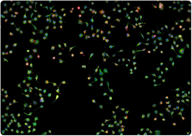
When the generation of photons by two or more different fluorescent markers overlaps during fluorescence microscopy, colocalization is said to have occurred. This is particularly useful in the diagnosis of chromosomal disorders and research into the role of specific genes.
Skip to:
 Damian Ryszawy | Shutterstock
Fluorescence microscopy is a commonly employed method of investigating the mechanisms of cellular function. Fluorescent molecules known as fluorophores are introduced to a sample, which become activated upon binding or coming into close contact with molecules of interest. This allows the presence and concentration of the target molecule to be inferred.
Antibodies may also be utilized for their highly specific binding with antigens, and the primary antibody can be labeled with a fluorophore. Alternatively, a secondary antibody that binds to the primary antibody may be labeled with the fluorophore, with each being added sequentially.
Why perform colocalization studies?
Colocalization studies are most useful in determining whether two or more biomolecules are affiliated with the same cellular structures. For example, an investigation into the purpose of two proteins may reveal that they are both involved in the function of the endosome.
Colocalization studies allow the exact location of cellular structures of interest to be determined, and features that they have in common can be examined quantitatively. The stages of transcription, signaling events, and cellular metabolism can be elucidated through colocalization studies.
If two probes spatially overlap in colocalization studies, then this is defined as a co-occurrence. If co-occurrence takes place throughout the cell, and the two probes are proportionately distributed, then correlation has occurred. For example, two fluorophores that interact with different areas of the same cellular component would be expected to correlate in this way.
Alternatively, if one fluorophore is intended to interact with a vesicle and a second to interact with the contents of the vesicle then they would co-occur, but not necessarily correlate, as each vesicle may contain multiple target molecules.
Colocalization protocol
Samples must firstly be prepared from cell cultures or cell or tissue excision from living animals or plants. The details of such preparations depend largely on the type and source of the sample, though ultimately they must adhere to glass slides that are suitable for microscopy.
Fluorophores or primary antibodies that bind with the molecule of interest must be selected carefully in order to avoid off-target binding. The approach of using primary antibodies followed by tagged secondary antibodies is highly supported by commercially available kits. Red and green fluorophores are frequently selected, appearing yellow when co-occurrence takes place.
Confocal microscopes are used to image cells that have been exposed to fluorophores. A laser is used as the light source that penetrates the sample to a specific depth, while a pinhole is used to prevent light that is out of focus from reaching the detector. This allows the sample to be imaged at multiple depths, and these two-dimensional images may be reconstructed to generate a three-dimensional image.
Computer software provided with appropriate algorithms then analyzes the image to determine the degree of co-occurrence and correlation that has taken place. The data collected by this software is frequently displayed as a scatterplot, comparing the intensity and frequency of one color on the x-axis with the intensity of frequency of the second fluorophore color on the y-axis.
Further Reading
Last Updated: May 22, 2019