Why Drug-Target Interactions Matter
What Is NMR Spectroscopy?
How NMR Detects Drug-Target Interactions
What Information Can NMR Reveal?
Advantages and Practical Limitations
Using Interaction Information to Guide Drug Discovery
Conclusion
References And Further Reading
Nuclear magnetic resonance (NMR) spectroscopy reveals how drug candidates interact with molecular targets, where binding affects the target, and how these interactions alter molecular structure and dynamics. These capabilities make NMR valuable for identifying weak binders, mapping binding sites, measuring affinity, and guiding drug discovery and lead optimization.
 Image credit: S. Singha/Shutterstock.com
Why Drug-Target Interactions Matter
Identifying a protein-drug interaction is an important step in drug discovery because binding can determine whether a candidate engages its intended target. However, binding alone does not establish therapeutic efficacy or functional inhibition. The structural and functional consequences of an interaction can depend on the target's folded state, post-translational modifications, and the structures adopted before and after ligand binding.1
Understanding these interactions can help researchers optimize compounds for stronger or more selective target engagement. In-cell NMR can further examine protein structure and drug interactions in living cells, providing information under conditions that more closely reflect the intracellular environment.1
What Is NMR Spectroscopy?
NMR spectroscopy provides information on molecular structure and interactions by measuring how atomic nuclei behave in a magnetic field. When placed in a strong magnetic field, NMR-active nuclei respond to radiofrequency pulses, producing signals that are processed into spectra.2
NMR is particularly useful for drug discovery because it can examine molecules in solution and provide atomic-level information about changes caused by ligand binding. When proteins are labeled with 15N or 13C, changes at individual amino acid residues can be followed by NMR. Binding of a ligand alters the chemical environment around affected residues, which can cause shifts in the corresponding NMR signals. These chemical shift perturbations (CSPs) can be monitored using experiments such as 1H-15N or 1H-13C heteronuclear single quantum coherence (HSQC) spectroscopy.3
Mapping these changes helps researchers locate regions affected by ligand binding and better understand how the ligand interacts with its target.3
How NMR Detects Drug-Target Interactions
NMR can detect binding using either ligand-observed or protein-observed experiments. Ligand-observed proton-based methods do not require an isotopically labeled target and can use relatively low protein concentrations. Binding can alter ligand relaxation rates, nuclear Overhauser effects (NOEs), diffusion coefficients, or spectral line widths, providing evidence of an interaction. Common ligand-observed approaches also include saturation transfer difference (STD) NMR, WaterLOGSY, and transferred NOESY.2,3
Protein-observed experiments can monitor changes in the resonances of individual residues after ligand addition. Chemical shift changes can identify regions affected by binding and, when interpreted alongside structural information, help define the binding interface. Comparing related ligands can also reveal residues important for recognition and differences in binding modes.3
However, ligand-induced perturbations may also arise from conformational changes transmitted away from the binding site; CSPs do not by themselves demonstrate direct ligand contact.3
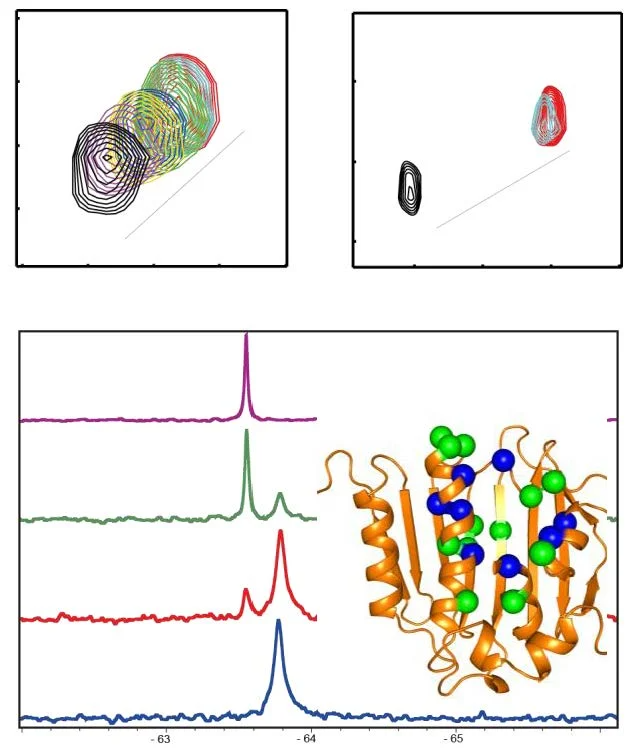 Protein-observed ¹9F NMR can distinguish changes in the conformational states of a G protein-coupled receptor (GPCR) in response to ligand binding. Changes in ¹9F resonances provide sensitive probes of the local environments of labeled sites and can reveal ligand-dependent conformational changes.³
NMR titration experiments can help quantify binding strength, including dissociation constants (KD). Results depend on the experimental design and binding model. NMR is also useful for studying weak interactions, making it valuable in fragment-based drug discovery. Protein-observed NMR can identify residues whose signals change when a ligand binds. Mapping these chemical shift perturbations onto a protein structure can help identify regions involved in binding and support structure-based drug design.2,3
However, a change in a residue's environment does not necessarily prove that the residue contacts the ligand directly, because conformational or allosteric changes can produce effects at more distant sites.3
NMR can reveal changes in molecular motion and conformational exchange after ligand binding. Different measurements provide complementary information. For example, the nuclear Overhauser effect (NOE) can indicate spatial proximity between interacting molecules, while relaxation measurements reveal local molecular motion.2,4
Diffusion measurements take a different approach by tracking translational movement and, under suitable experimental conditions, can help estimate how much of a drug is bound. In systems where free and bound species exchange rapidly on the diffusion timescale, an observed diffusion coefficient can reflect a population-weighted contribution from the different states, allowing the bound fraction to be estimated with an appropriate model.4
These approaches can therefore provide a broader picture of how an interaction affects molecular behavior. 19F-NMR can also monitor changes in the local environment of fluorinated probes and has been used to investigate conformational changes in G protein-coupled receptors.2,3
Comparative NMR experiments using related compounds can help assess differences in binding behavior and identify structural features associated with stronger interactions. Such information can guide medicinal chemistry and lead optimization.3
Advantages and Practical Limitations
A major advantage of NMR is its ability to detect weak interactions. Ligand-observed approaches can avoid isotope labeling of the target and are useful for identifying weak-affinity ligands, including fragments with micromolar to millimolar binding affinities. Other experiments, particularly protein-observed multidimensional methods, may require isotopically labeled proteins and suitable spectral quality.2,3
Molecular motion and transient interactions can also be examined by NMR when the experimental conditions and exchange behavior permit their detection. Along with the structural information obtained by X-ray crystallography and cryo-electron microscopy (cryo-EM), this approach offers a view of changes in molecular behavior. Because the measurements are performed in solution, NMR can also capture aspects of molecular dynamics that may be difficult to discern from a static structure.2
However, NMR has practical limitations. Several factors influence how readily NMR can be applied, including sensitivity, sample quantity, protein stability, spectral quality, and target size. For protein-observed experiments, isotopic labeling of the protein may also be necessary. Conventional solution NMR becomes increasingly challenging for larger proteins because slower molecular tumbling broadens resonances and reduces spectral sensitivity, although specialized labeling and experimental strategies can extend the accessible molecular-weight range.2,3
The practical challenges become greater with membrane proteins and other difficult targets, which can be hard to express and purify and may require careful handling to remain suitable for analysis. Spectral overlap and incomplete resonance assignments can also complicate interpretation. Multidimensional experiments used for resonance assignment can require acquisition periods ranging from minutes to days, so maintaining sample stability throughout data collection is important. Therefore, no single NMR experiment provides every type of information for every target, and complementary techniques are often needed.2,3
NMR can contribute throughout hit identification and lead optimization. In fragment-based drug discovery, ligand- or protein-observed NMR can identify weak binders that may serve as starting points for more potent compounds. Protein-observed approaches can then help determine where fragments bind and guide strategies for linking or optimizing them. NMR can also be used to validate hits obtained from high-throughput screening and can help reduce false-positive assignments by directly examining protein-ligand interactions.3
The development of KRAS inhibitors illustrates how NMR can reveal druggable features that may not be apparent from a single static structure. NMR studies of Kirsten rat sarcoma viral oncogene homolog (KRAS) identified multiple rapidly interconverting conformational states and helped reveal a surface pocket associated with one of these states. Subsequent fragment screening and complementary structural studies contributed to the development of inhibitors targeting this region.2
Another example is structure-activity relationship (SAR) by NMR, in which protein-observed experiments are used to identify fragments that bind at neighboring regions of a target. In studies of B-cell lymphoma 2 (Bcl-2) family proteins, NMR helped identify fragment binding sites and informed a strategy for linking fragments into more potent molecules. Venetoclax, an approved Bcl-2 inhibitor, was developed using this fragment-based strategy. NMR can also support hit validation and mechanistic studies by confirming target engagement, comparing binding affinities, mapping binding sites, and examining conformational changes.1,2
Conclusion
NMR spectroscopy provides complementary information about drug-target interactions, including whether a compound binds, how strongly it binds, where binding affects the target, and how the interaction changes molecular structure and dynamics. Its ability to study weak interactions, molecular motion, and solution-state behavior makes it valuable alongside X-ray crystallography, cryo-EM, and biochemical or biophysical assays.2,3
Rather than serving as a standalone measure of therapeutic activity, NMR helps build a more detailed picture of target engagement and molecular mechanism. By combining ligand-observed, protein-observed, and, increasingly, in-cell measurements, NMR can provide complementary views of molecular recognition from initial hit identification through lead optimization and studies of interactions in a cellular environment.1,2,3
References And Further Reading
- Washington, J.D., Burz, D. S., and Shekhtman, A. (2013). CHAPTER 4: In‐Cell NMR Spectroscopy to Study Protein–Drug Interactions, New Applications of NMR in Drug Discovery and Development, ed. L. Garrido and N. Beckmann, The Royal Society of Chemistry, pp. 134-161, DOI: 10.1039/9781849737661-00134, https://books.rsc.org/books/edited-volume/1007/chapter-abstract/803409/In-Cell-NMR-Spectroscopy-to-Study-Protein-Drug
- Caceres-Cortes, J., Falk, B., Mueller, L., Murali Dhar, T G. (2024). Perspectives on Nuclear Magnetic Resonance Spectroscopy in Drug Discovery Research. J. Med. Chem., 67 (3): 1701–1733. DOI: 10.1021/acs.jmedchem.3c02389, https://pubs.acs.org/doi/10.1021/acs.jmedchem.3c02389
- Li, Y., & Kang, C. (2017). Solution NMR Spectroscopy in Target-Based Drug Discovery. Molecules, 22(9), 1399. DOI: 10.3390/molecules22091399, https://www.mdpi.com/1420-3049/22/9/1399
- Kumar, B. P. (2025). NMR Approaches to Study Drug–Surfactant Interactions: Insights From NOE, Relaxometry and Diffusometry, Magnetic Resonance in Chemistry, 63, 11:994–1015, DOI: 10.1002/mrc.70030, https://analyticalsciencejournals.onlinelibrary.wiley.com/doi/10.1002/mrc.70030
Last Updated: Oct 8, 2026