Introduction: Beyond Binding Studies
Why Fluorine is Attractive for Drug Discovery
Monitoring Enzyme Reactions with Fluorine NMR
Benchtop NMR Systems
Case study: Inhibitor Screening with Benchtop 19F NMR
Advantages and Limitations of Benchtop NMR
Future Directions
References and Further Reading
Fluorine-19 NMR provides a powerful approach to evaluating enzyme inhibitors, combining direct biochemical measurements with applications in drug discovery. Recent advances in benchtop spectrometers demonstrate its potential for accessible, quantitative inhibitor screening while highlighting practical limitations in sensitivity, resolution, and throughput.
 Image credit: remotevfx.com/Shutterstock.com
Fluorine is rare in most biological samples but abundant in many modern drugs. This difference has proved important in drug discovery, where Fluorine-19 Nuclear Magnetic Resonance (fluorine NMR) is used to study how candidate compounds interact with their targets.1,2
Introduction: Beyond Binding Studies
NMR has long been used in drug discovery to detect whether a small molecule attaches to a protein target. Attachment alone, however, says little about activity, since binding does not always mean inhibition. Fluorine NMR goes further by measuring enzyme activity when a suitable fluorinated substrate or cofactor is available.
A substrate carrying a fluorinated group gives a signal that moves when the enzyme converts it, so the reaction, and its suppression by an inhibitor, can be read from one spectrum. This functional approach, known as n-FABS (n-Fluorine Atoms for Biochemical Screening), differs from ligand-observed and protein-observed fluorine NMR experiments that primarily report molecular binding or conformational changes.1-4
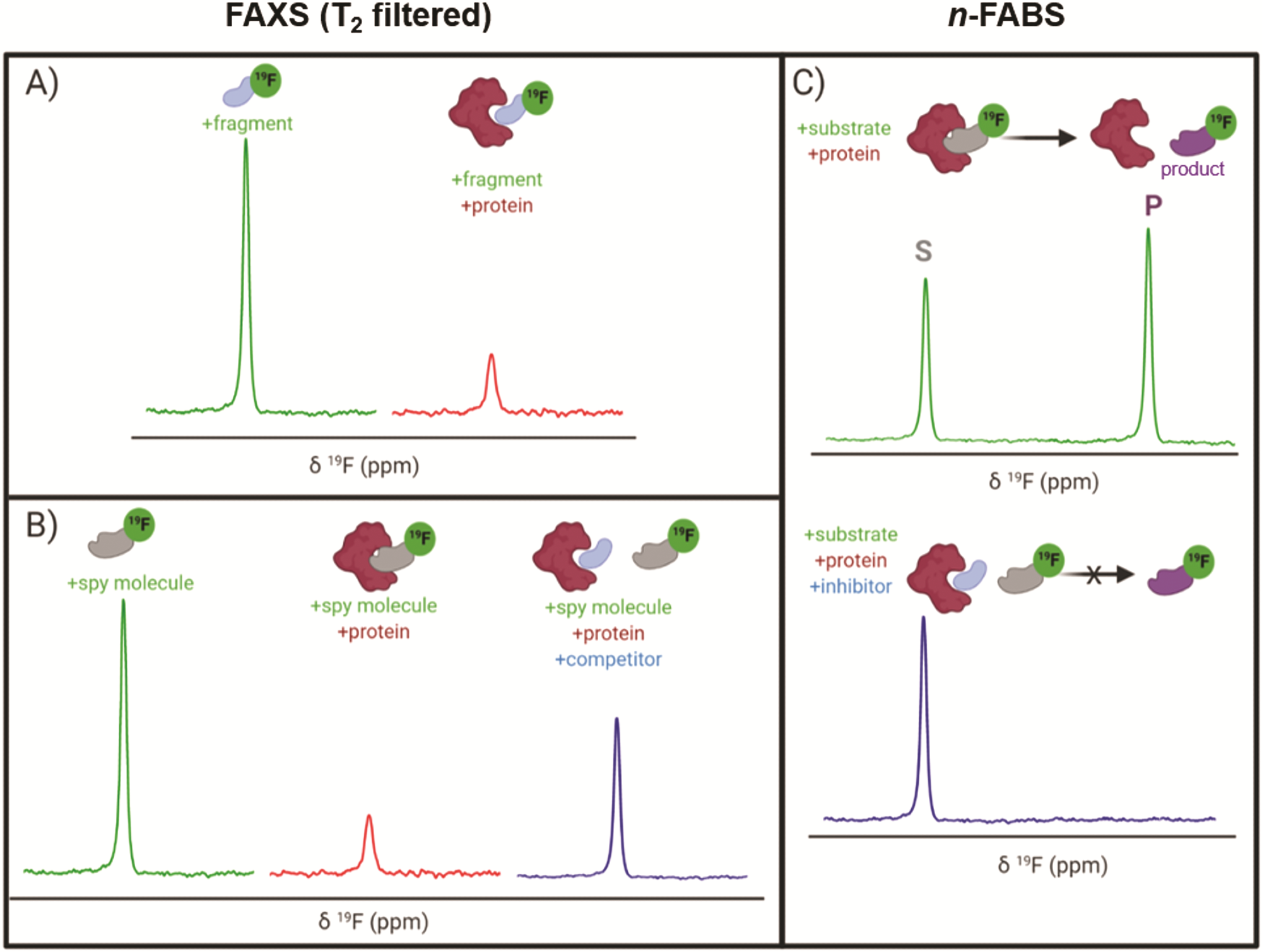 Fluorine-19 NMR approaches to drug discovery. (A) Fragment binding changes the fluorine NMR signal. (B) Competitive binding restores the signal of a displaced fluorinated reporter molecule. (C) Functional n-FABS assays distinguish substrate and product signals, allowing enzyme activity and its inhibition to be measured. Image cred: Buchholz and Pomerantz (2021).
Why Fluorine is Attractive for Drug Discovery
Fluorine NMR detects fluorine-19, which occurs naturally as a single isotope and responds to a magnetic field with about 83% of hydrogen's intrinsic NMR receptivity. Its signals react strongly to the surrounding chemical environment, so even a modest change near a fluorinated group can visibly displace them. Fluorine signals also span several hundred ppm as compared to about 14 ppm for hydrogen. Therefore, peaks that would overlap in a proton spectrum can often be resolved in fluorine NMR.1,4,5
Proteins and most biological media contain almost no fluorine, so fluorinated molecules are easy to detect against the absence of a biological background signal. This allows screening in conditions resembling a cell's interior, culture medium, or even blood, although the feasibility of measurements in complex biological media depends on probe stability, sample composition, and spectral quality. Fluorine NMR can also help distinguish specific binding from certain nonspecific interactions. One study reported that up to 30 compounds have been measured together at high field.2,3,5
Fluorine is also common in pharmaceutical products. A 2007 analysis found that about one in five drugs contained fluorine, and later tallies put the share of newly approved small-molecule drugs containing fluorine at 29% to 47% in individual years. Many drug-like molecules therefore already carry a usable NMR probe, although fluorine-containing drugs are not necessarily suitable substrates for functional enzyme assays.1,2
Monitoring Enzyme Reactions with Fluorine NMR
In fluorine NMR assays, a substrate is tagged with a fluorine tag, often a trifluoromethyl (CF3) group whose three equivalent fluorines give one strong, sharp signal. The tag need not sit at the reactive site. The enzyme converts the substrate into a product that resonates at a different position; with both appearing in one spectrum, their integral ratio gives the fraction converted when the signals are sufficiently resolved and measured under quantitative conditions.1,4,5
Inhibitors of enzymatic reactions slow the conversion of the substrate to the product. Therefore, at a fixed time, the integrals give the percentage of inhibition based on the concentration of the remaining substrate. Detectability depends on the inhibition mechanism and substrate concentration. Furthermore, the partial inhibition at one concentration also provides a rough estimate of the half-maximal inhibitory concentration or IC50 value, although reliable IC50 determination requires measurements across multiple inhibitor concentrations.4
However, the IC50 value depends on assay conditions, such as inhibitor and substrate concentration, and should not be treated as interchangeable with binding affinity or an inhibition constant. Furthermore, the duration for which the enzymatic reaction is run can also impact the IC50 value, as greater substrate turnover percentages can lead to non-linear enzymatic reaction behavior and higher IC50 values.4
Other biochemical assay methods present their own limitations. Radioactive labeling requires product separation, while direct binding NMR needs milligram quantities of labeled protein, and competition-based screening requires a reporter ligand.2,6
Fluorine NMR can also monitor enzyme activity using fluorinated cofactors. In one study, fluorinated analogs of phosphoadenosine 5′-phosphosulfate (PAPS) enabled continuous monitoring of sulfotransferase activity and screening of potential inhibitors, demonstrating the method's applicability beyond conventional fluorinated substrates.6
Benchtop NMR Systems
Benchtop NMR spectrometers have become commercially available in the last decade. These use permanent magnets at 60 to 90 MHz and do not require cryogenic magnet cooling, unlike conventional high-field superconducting spectrometers. However, their sensitivity is substantially lower, with reported differences of approximately 150–400-fold for certain instrument and probe comparisons, and their frequency resolution in hertz is reduced at lower magnetic field strengths. They also commonly use sample volumes of 300 to 600 µL, as compared to the 20 µL sample volumes achievable with certain high-field microprobe configurations.3
Benchtop NMR spectrometers offer advantages in price and ease of use. They cost about $50,000 to $200,000, as compared to the $1–$1.5 million required for a cryoprobe-equipped 600 MHz system. Benchtop units can also be installed in many conventional laboratories without the infrastructure needed for superconducting magnets and used by non-specialists.3
Hyperpolarization methods such as Signal Amplification by Reversible Exchange (SABRE) and Photochemically Induced Dynamic Nuclear Polarization (photo-CIDNP) are now being explored to offset low sensitivity, and fluorine detection is used to improve spectral discrimination through its broad chemical-shift dispersion. These hyperpolarization approaches are promising but are not yet universal solutions for routine fluorine-based enzyme inhibition measurements.3
Case study: Inhibitor Screening with Benchtop 19F NMR
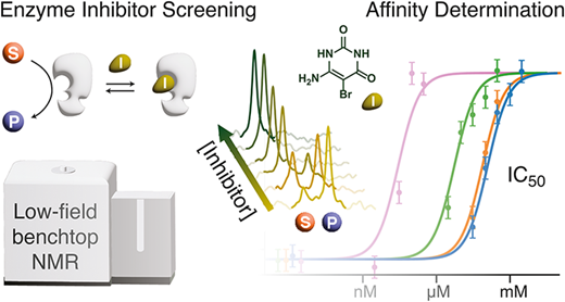 Benchtop fluorine-19 NMR enables enzyme inhibitor screening by measuring changes in substrate and product signals and determining inhibitor potency from IC50 concentration–response curves. Image credit: Jordan et al. (2026)
In a 2026 Analytical Chemistry study, a team of European researchers showed that an inexpensive benchtop NMR spectrometer can rank enzyme inhibitors. They assayed thymidine phosphorylase, using a bacterial enzyme as the experimental model, and measured the conversion of trifluorothymidine into trifluorothymine.4
They ran the enzymatic reaction in the presence and absence of different inhibitors at 50 µM concentration, and determined the IC50 values for each inhibitor. The reactions were run for two hours at 37 °C in 500 µL of buffer with 1 mM substrate. Fluorine signals from substrate and product, separated by 12.5 Hz on an 80 MHz instrument, were integrated directly.4
At 50 µM, each inhibitor took about 25 minutes of NMR measurement time. Two inhibitors blocked the reaction completely, one did nothing, and two partially inhibited it. The dose-response series estimated IC50 values ranging from 29 nM to 154 µM. Two of the values matched published figures for the human thymidine phosphorylase enzyme.4
The study presented some limitations. The assay required about 30 minutes of measurement time per inhibitor, and the IC50 calculation took about three hours, which does not make the assay high-throughput. While a paramagnetic additive was used to cut measurement time roughly fourfold, it also caused some additional line broadening and reduced spectral separation, potentially compromising accurate peak integration.4
Advantages and Limitations of Benchtop NMR
In benchtop NMR instruments, substrate conversion is measured directly, so the readout reflects enzyme activity rather than a coupled signal. A suitably fluorinated substrate can serve as its own reporter without requiring a separate optical probe. Quantification is reliable when acquisition and analysis conditions are controlled. One major advantage is that equipment costs fall well below those of high-field systems, though capabilities differ and have limitations.3,4
The major limitations include lower sensitivity and poorer frequency resolution than high-field NMR spectrometers, and potential overlaps affecting quantification. Benchtop NMR throughput is modest, as shown in the case study. Furthermore, substrates must carry fluorine, and that change may alter enzyme behavior. Each assay needs its own validation of conditions and measurement requirements, and the method also requires larger sample sizes. Additional practical considerations include substrate solubility, fluorinated-probe stability, relaxation behavior, inhibitor solubility, and possible interference with the enzyme or the NMR measurement itself.2,4,6
Fluorine NMR measurements of labeled proteins present a different set of challenges. Chemical-shift anisotropy can broaden signals, particularly for large biomolecules at high magnetic fields. Approaches involving mobile CF3 labels or fluorine-carbon TROSY experiments can help address these limitations, but they should not be confused with the direct substrate-conversion measurements used in functional benchtop assays.1,5
Future Directions
Fluorinated-probe technology continues to expand, with larger libraries and in-cell uses being reported. Benchtop instruments are being connected to flow-through setups, and one study reported a 96-well reaction format used in the sulfotransferase screen, which suggests routes toward automation. However, preparing reactions in multiwell plates does not by itself establish high-throughput NMR acquisition, since spectral measurement time and sample handling can remain limiting factors.2-4,6
Growing evidence on the efficacy of fluorine NMR in exploring biomolecule-ligand interactions also indicates that it will play a crucial role in fragment screening as a part of Fragment-Based Drug Discovery.7
Further progress is likely to depend on improved fluorinated substrate and cofactor design, more efficient sample handling, automated spectral analysis, and approaches that increase sensitivity without sacrificing quantitative accuracy. Combining these advances with lower-cost instruments could broaden access to functional NMR assays, particularly where conventional optical or coupled biochemical methods are unsuitable.3,4,6,7
References and Further Reading
- Werle, Y., & Kovermann, M. (2024). Fluorine labeling and 19F NMR spectroscopy to study biological molecules and molecular complexes. Chemistry – A European Journal, 31(2). DOI:10.1002/chem.202402820, https://chemistry-europe.onlinelibrary.wiley.com/doi/10.1002/chem.202402820
- Buchholz, C. R., & Pomerantz, W. C. K. (2021). 19F NMR viewed through two different lenses: ligand-observed and protein-observed 19F NMR applications for fragment-based drug discovery. RSC Chemical Biology, 2, 1312. DOI:10.1039/D1CB00085C, https://pubs.rsc.org/en/content/articlehtml/2021/cb/d1cb00085c
- Bütikofer, M., Stadler, G. R., & Torres, F. (2024). Rescaling NMR for a larger deployment in drug discovery: Hyperpolarization and benchtop NMR as potential game-changers. Chemistry–Methods, 4, e202400009. DOI:10.1002/cmtd.202400009, https://chemistry-europe.onlinelibrary.wiley.com/doi/10.1002/cmtd.202400009
- Jordan, C., Piotto, M., Loss, S., Dalvit, C., & Gossert, A. D. (2026). 19F NMR functional screening on a benchtop NMR instrument: Theoretical analysis and efficient application to drug discovery. Analytical Chemistry, 98, 14049–14059. DOI:10.1021/acs.analchem.5c07368, https://pubs.acs.org/doi/10.1021/acs.analchem.5c07368
- Boeszoermenyi, A., Ogórek, B., Jain, A., Arthanari, H., & Wagner, G. (2020). The precious fluorine on the ring: Fluorine NMR for biological systems. Journal of Biomolecular NMR, 74, 365–379. DOI:10.1007/s10858-020-00331-z, https://link.springer.com/article/10.1007/s10858-020-00331-z
- Mlynarska-Cieslak, A., Chrominski, M., Spiewla, T., Baranowski, M. R., Bednarczyk, M., Jemielity, J., & Kowalska, J. (2022). Fluorinated phosphoadenosine 5′-phosphosulfate analogues for continuous sulfotransferase activity monitoring and inhibitor screening by 19F NMR spectroscopy. ACS Chemical Biology, 17, 661–669. DOI:10.1021/acschembio.1c00978, https://pubs.acs.org/doi/10.1021/acschembio.1c00978
- Li, Q., & Kang, C. (2024). Perspectives on applications of 19F-NMR in fragment-based drug discovery. Molecules, 29, 5748. DOI:10.3390/molecules29235748, https://www.mdpi.com/1420-3049/29/23/5748
Last Updated: Oct 8, 2026