An interview with Dr Martin Groher conducted by Alina Shrourou, BSc
Please give an overview of digital pathology today, and how pathologists currently share histology images to discuss diagnoses and further their studies.
There are two problems that pathologists face today. The first is, that a pathologist’s diagnosis using tissue sections is becoming more complex. There are many more biomarkers, and clinicians now ask for a more precise diagnosis. Secondly, the number of pathologists worldwide is declining every day, as the profession is often overlooked in favor of other specializations.
There is an opportunity for digital pathology to help solve these problems through telepathology. This is where workloads can be distributed remotely, and you can better respond to high work peaks.
Furthermore, digital pathology enables image analysis, which can optimize tasks during the diagnostic process for the pathologist and significantly speed up the diagnosis. Experts believe that every pathology lab needs to switch to digital pathology in the next three to five years to stay competitive.
How do the current digital pathology systems produce barriers, especially in smaller labs?
Digital pathology usually comes at high costs and effort, so you will have to invest quite significantly to get your lab to produce digital pathology outcomes. Currently, full-fledged digital pathology solutions cost around 0.5 to 1.5 million US dollars.
You will need a virtual slide scanner, storage systems, software for workflow management etc. Then, you must train all your employees which requires more costs. So, in the beginning, this solution is not cheap and takes quite some years to amortize. Unfortunately, many small labs cannot afford this.
Today, there are only few labs in the world that have gone completely digital. Some countries, like the Netherlands, or Scandinavian countries, are advanced in this area; but, for instance in Germany, or in the US, the transition to digital pathology in the clinical field is really slow.
With hardly any sources to get informed and without low-cost solutions to get started slowly, labs have a hard time anticipating or experiencing the benefits of digital pathology. This is the main barrier.
What methods are there for pathologists to share and discuss histology images? What advantages does collaborating online bring to them?
Until the middle of 2017, there was no dedicated low-cost solution available for sharing and discussing histology images. Pathologists therefore turned toward ‘off the shelf’ tools like email, WhatsApp, Dropbox, or other file sharing systems, to send their image data to colleagues for case discussion. They even use social media networks such as Twitter and Facebook.
Pathologists are taking a risk by sharing health information via these unspecific communication channels, but they do this because there are significant advantages.
First of all, pathologists do not have to send the tissue or section by post to get a second opinion. Physical sending not only takes a long time but also bears the risk of loss – in the case of the glass slide you may even lose the evidence on which the diagnosis was made.
Digitizing pathology findings and sharing images online also provides better freedom to include a wider range of partners to discuss results. The pathologist can choose an expert (or several) with a high degree of specialism anywhere in the world to discuss his or her results with, rather than being limited to the local area.
One further advantage is that the pathologist could directly engage with patients, if needed. Image sharing was not designed for pathologists to talk to patients, but in a world where everyone can get informed through the internet, patients will often want to double-check their diagnosis with their pathologist to understand.
Therefore, these tools for online sharing and discussion give the opportunity to engage with other pathologists and patients, too.
Do the patients who have their images shared remain anonymous?
The pathologist must make sure that everything is de-identified and properly anonymized when sharing images – this is true for any image used in medical publication and on image sharing platforms. But of course, humans do make mistakes.
This brings us back to the point that there is a need for a dedicated solution – to take care of all of this and to avoid the sharing of protected health information through unprotected channels.
How easy or difficult is it for pathologists outside of large labs and research centers or those in remote regions, to access, share and discuss pathology images?
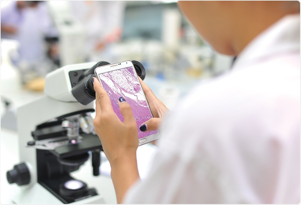
Small or mid-sized labs do usually not have access to any digital pathology solutions. They work on microscopes that will be equipped with digital cameras, or they will take photos through the ocular with their smart phones.
However, sharing and discussion is challenging, because free or low-cost communication channels are not there, or they are not safe. Therefore, it is definitely more difficult for pathologists outside of large labs to access, share and discuss pathology images.
The access to high quality image data, especially for those pathologists who are working in less developed countries and do not have advanced equipment, is also limited. Very often, digital pathology systems in large labs are incompatible for remote use.
This is because they are usually missing easy to access components to view and collaborate on data remotely, and the partnering lab would have to invest into software and integrate it, too.
There are a couple of web platforms which host high quality images, and make these available for the public, or for user restricted groups. These web platforms are focused on educational purposes and are often homemade by university hospitals or large institutions – and many lack the large variety that is out there for pathology images.
Please outline the new cloud-based image sharing platform that microDimensions has developed? What was the motivation and vision?
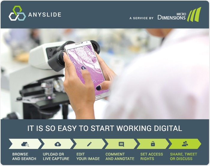
This is really where we step in with Anyslide. Here, our goal is to empower pathologists to benefit from digital pathology, no matter how advanced they are, or their lab is, on the digital journey. We want to compile and provide access to the largest variety of digital pathology data from any region and at the same time enable online collaboration on images – publicly or privately, and always securely.
Anyslide is also very easy and intuitive to use, and of course, by providing a free, browser-based platform we eliminate the need for a pathologist to first ask for budget and then ask IT staff to get started.
The crucial information for all the pathologists and the pathology diagnosis lies in the images. Anyslide provides the easiest access to this information: via an image-hosting platform including tools for case discussion, sharing, and management.
Anyslide - Digital Pathology Now - Collaboration
How is the Anyslide platform unique compared to other existing platforms?
Anyslide is driven by digital pathology professionals and dedicated to pathologists.
Firstly, the platform is fully web and browser based. You can use it without any installation or expensive set up. The main difference is that it’s dedicated to pathologists, not for pathology labs.
This means that we avoid the costly effort to integrate with legacy systems, like laboratory information systems. By saving this effort and maintenance we cut down costs and are able to provide the first professional digital pathology platform entirely free of charge.
It is also different to “home-made” image collaboration platforms established by individuals, universities or other organizations. Anyslide provides an intuitive and user-proven interface as well as professional maintenance.
Finally, it is different to solutions from digital pathology hardware vendors, as it is truly neutral to the underlying imaging method - no matter if a lab has a high-end virtual slide scanner (from any vendor), a camera mounted on a microscope, or simply some smartphone snapshots through the ocular, Anyslide supports image-based collaboration.
What type of images and information can be shared on the platform? Are you able to search through the database for specific tissue types or conditions?
Anyslide users can share digital images created on their existing set-up. These include static images like jpeg, tiff or png (as taken by live capturing from microscope cameras) as well as whole slide images (WSI). This approach opens up digital pathology to a much larger group of pathologists than ever before.
Anyslide sets no limits to the information images are attributed with. Users can add tissue types, staining, condition, or diagnosis to their uploads. We clearly encourage and foster this diversity. The database can be searched by keywords and tags (#-based search) and a case-based organization of images is one of our next immediate steps for new functionality.
In the future, Anyslide may also offer methods for image analysis, to e.g. automatically tag them or compute a diagnostic score.
What happens if pathologists upload something with an incorrect diagnosis? Is there a proof-checking system in place?
This is something which is very important. We validate everything in a double, or triple fashion - by letting the crowd vote. When a new image is uploaded, pathologists on system that work within the expertise relevant to the recently uploaded image, will be asked if they agree with the diagnosis, set staining etc. Then with that, we create a vote and we can tell whether it's correct, or not.
Who can access and use Anyslide? Is the platform open access or is there a cost attached to using the tool?
Everyone can access Anyslide. You can put your images online and share them publicly, and then you can view them, whether you're a user of Anyslide or not. All you need is the link!
Of course, in order to edit, annotate, and describe your images, you need to have an account because we want to avoid an anonymous person annotating images. Account registration is free of charge.
For each image that you upload, you can choose to restrict it from the access of others, and just share it with a certain group of people for case discussions, for example; or you can also share it with the public for educational purposes.
You can also post a public image or a snapshot of an area of interest directly on your Twitter account from within Anyslide – for Whole Slide Images (WSI) this holds the possibility that your Twitter followers can follow back to the platform and to view it in its entirety and detail, add annotations, comments and questions.
Sign-up and the use of Anyslide is, and will stay, free of charge. We offer a professional subscription plan for those users that upload images exceeding a certain storage limit (currently, this is 5 GB per user). This small monthly fee will cover the larger storage costs.
What limitations are there to the Anyslide platform?
We launched Anyslide just two months ago, at the beginning of September 2017. That means that the platform is very young and the potential for future functionality and use cases is immense.
The nice thing is that it's an opportunity for pathologists to get involved early and give us feedback. At this starting stage we are both eager and able to process feedback on user experience and new use cases and functionality.
What do you think the future holds for histology image collaboration through sharing platforms such as Anyslide in the healthcare and the pharma industry?
We want Anyslide to become the hub for pathology experts of all sub specialties worldwide. This should also include possibilities for every pathologist to offer their expertise to contractors from pharma or healthcare industries.
Currently, everyone is talking about how automation, computational analysis and artificial intelligence (AI) can revolutionize outcomes in the medical and pharma domain.
The problem that AI algorithms have is that they need a very large training and testing data base to be able to produce correct results. With the vision to become the world’s largest database of curated (=labeled) pathology image data, Anyslide will help to provide these rich data sets to companies working on better decision algorithms for oncology, for instance, or any other field of pathology.
Where can readers find more information?
Further information can be found on Anyslide.com
About Dr. Martin Groher 
Dr. Martin Groher is CEO and co-founder of microDimensions, and he is responsible for strategy, sales, marketing, innovation and change management.
He received his PhD degree in Computer Science at the Chair for Computer Aided Medical Procedures, Department of Informatics at the Technical University of Munich.
After this, he led a group of scientists at the Technical University of Munich to work on histology reconstruction, where the original idea to microDimensions was born.
His visionary thinking combined with his deep knowledge of the digital pathology market and his profound expertise in image processing and analysis guarantee the continuous success of microDimensions' products and services.
 microDimensions launches new version of high precision whole slide alignment software