An interview with David Jäckel, PhD, conducted at SfN 2018 by Alina Shrourou, BSc.
Why is it valuable to stimulate electrogenic cells in vitro for neuroscience research?
One reason selective stimulation is valuable is that it allows you to control the activity of certain cells rather than just recording their spontaneous activity. You can thus not only measure, but also interact with your cells.
Secondly, one can investigate neuronal excitability. How much voltage is needed to evoke an action potential? This biologically relevant number can tell us important information about the maturity and health of cells.
Electrogenic cells can be precisely stimulated with our microelectrode array MaxOne, as every neuron is always surrounded by multiple electrodes.
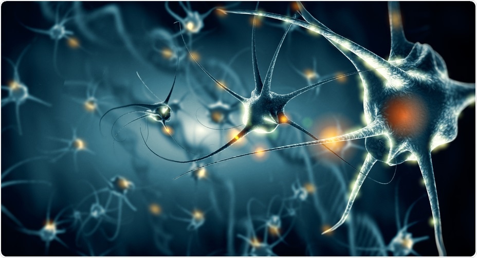 © Giovanni Cancemi/Shutterstock.com
How can CMOS be used to enhance HD-MEA systems?
CMOS stands for complementary metal-oxide semiconductor, which is a standardized technology used in every cell phone and every computer. We use this technology to build state-of-the-art microelectrode arrays with tens of thousands of electrodes and readout circuitry integrated in a small chip.
How does MaxWell Biosystems’ MaxOne provide whole sample electrical imaging?
Our system features 26,400 electrodes at very high density (3,265 electrodes/mm2), which means that there is always an electrode close to a neuron that can detect its activity. Thus, by scanning through the 26,400 electrodes using 1,024 read-out channels at a time, a whole-sample electrical image is acquired. Depending on the desired imaging resolution, this automated scan through the whole array requires between 5 and 20 minutes.
The feature of selecting electrodes is very helpful because you usually only want to measure where you have cells. By selecting specific electrodes, you can adapt to your sample and optimize the information obtained from the recordings.
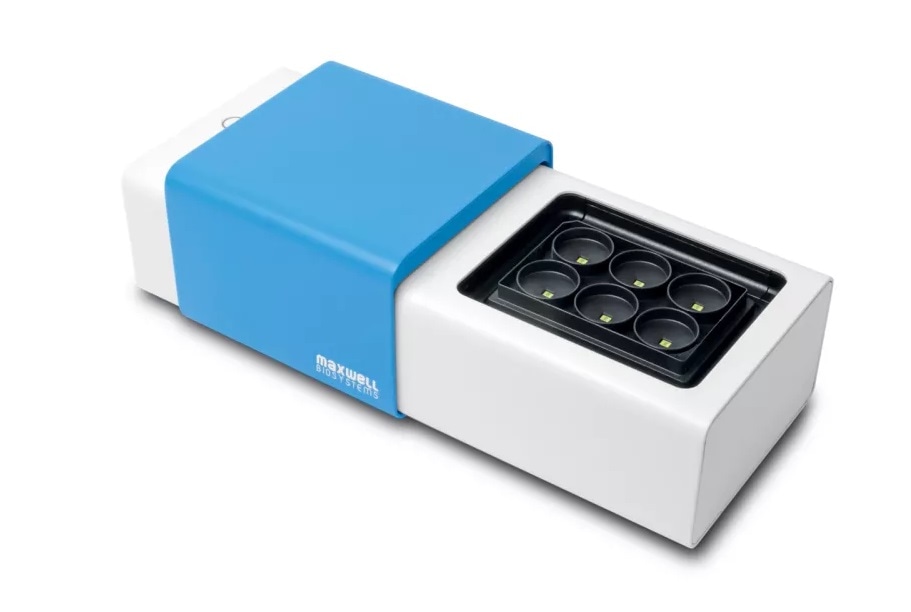
What data can be collected by using the high-throughput recordings made by the MaxTwo?
MaxTwo allows to parallelize the experiments using 6 wells at a time. The different types of data, that can be recorded, are equal to the ones with our single-well device MaxOne.
One type is what we call the “activity map”, which is an image of how the cells on the array fire and where they are positioned.
The second type of data is called “network activity”, which involves selecting electrodes with high activity and then recording them simultaneously. This data can be used to analyze the interaction between cells, such as how much they are connected, their synchronicity, and their bursting activity.
Finally, the third type of data is one of the unique features of our system. After identifying individual cells, we record their signals with hundreds of electrodes. Using this, we can access subcellular features, such as signals around the somatic and the dendritic area and signals propagating along the axons. This allows to precisely measure axonal conduction velocity, which is very challenging to achieve using alternative techniques.
What makes the MaxOne and MaxTwo differ from other high-resolution functional imaging solutions on the market?
There are three points to this. First, we have the largest number of electrodes; 26,400 electrodes on an area of 2x4 mm2. It's also the largest sensing area.
Secondly, the distance between two neighbouring electrodes is 17.5 micrometers so we have, by far, the highest resolution available.
The third point is the signal-to-noise ratio, which is in the order of 4 microvolt RMS during experiments. This means that we can measure very small signals with our technology. Signals propagating along the axon of a neuron, are typically very small. Therefore, a high signal to noise ratio is a key to measuring and analyzing the axonal conduction velocity.
About David Jäckel, Ph.D.
.jpg)
After finishing a Masters of Science in Electrical Engineering at ETH Zurich in 2008, David went on to complete a PhD in Neuroscience there in 2014.
Following this, David became a consultant at AWK for the next 3 years, before moving onto MaxWell Biosystems in 2018, where he holds the position of Product and Project Manager.