Stem cell models of the retina and optical nerve have been used to identify previously unknown genetic markers of glaucoma, in research jointly led by scientists from the Garvan Institute of Medical Research, the University of Melbourne, and the Centre for Eye Research Australia. The findings open the door to new treatment for glaucoma, which is the world's leading cause of permanent blindness.
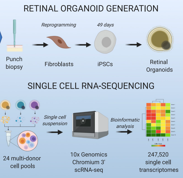
 The study used skin samples to generate stem cell models, and then carried out detailed single-cell genetic sequencing. Image Credit: Garvan Institute
We saw how the genetic causes of glaucoma act in single cells, and how they vary in different people. Current treatments can only slow the loss of vision, but this understanding is the first step towards drugs that target individual cell types."
Joseph Powell, Joint Lead Author and Professor, Garvan Institute of Medical Research
The research, published today in the journal Cell Genomics, comes out of a long-running collaboration between Australian medical research centers to use stem-cell models to uncover the underlying genetic causes of complicated diseases.
Glaucoma damages cells in the optic nerve, the part of the eye that receives light and sends it to the brain. It is impossible to take samples from this part of the eye in a non-invasive way, which limits research.
Instead, to create samples for the study, researchers took skin biopsies from participants with and without glaucoma. The skin cells were reprogrammed to revert to stem cells, and then guided into becoming retinal cells.
With 110 successfully converted samples, researchers sequenced more than 200,000 individual cells to generate 'molecular signatures'. Comparing signatures with and without glaucoma revealed key genetic components that control how the disease attacks the retina.
Studying glaucoma inside retinal cells created a 'context-specific profile' of the extremely complicated disease, says co-lead author Professor Pébay.
"We wanted to see how glaucoma acts in retinal cells specifically - rather than in a blood sample, for instance - so we can identify the key genetic mechanisms to target. Equally, we need to know which genetic variations are healthy and normal, so we can exclude them from a treatment," she says.
Across both healthy and diseased samples, researchers identified 312 genetic variants associated with the target retinal cells. Further analysis found 97 genetic clusters linked to the damage caused by glaucoma.
The study's third lead author, Professor Alex Hewitt, says the findings lay the foundation for research into new glaucoma treatments:
"Not only can scientists develop more tailored drugs, but we could potentially use the stem-cell models to test hundreds of drugs in pre-clinical assays. This method could also be used to assess drug efficacy in a personalized manner to assess whether a glaucoma treatment would be effective for a specific patient."
Source:
Journal reference:
Daniszewski, M., et al. (2022) Retinal ganglion cell-specific genetic regulation in primary open-angle glaucoma. Cell Genomics. doi.org/10.1016/j.xgen.2022.100142