TRM are distinguished by the expression of CD103, CD69, and CD49a. TRM plays a central role in the development of and the efficacy of cancer vaccines and has been related to improved prognosis and survival in a number of cancers. However, comparisons of TRM and cytotoxic CD8 T cells in the tumor microenvironment are limited.
What was the main objective of your study?
We set out to develop the HISTOPROFILE®-TRM multiplex panel to phenotype memory resident T cells in solid tumors and show the panel’s potential to study TRM subpopulations in NSCLC FFPE tissue resections. This was achieved using panel design and validation.
What were your main targets during the research?
The targets were CD3/CD8/CD49a/CD69/CD103; memory resident T cells – CD8+/CD103+; and subtypes of memory resident T cells – CD8+/CD49a+, CD8+/CD69+, CD8+/CD103+/CD69+, CD8+/CD103+/CD69+/CD49a+.
To achieve this, we sourced three NSCLC tissue resections samples from the Cerba Reseach Montpellier Biobank. The NSCLC tumor area on hematoxylin and eosin-stained slides was identified by a pathologist.
What application methods were applied during the validation of the study?
Once the slides had been identified, a sequential multiplex protocol with Opal® (Akoya Biosciences) fluorophores were applied on the BOND RX® (Leica) slide stainer. Following this, multispectral images were captured using the VECTRA® PolarisTM (Akoya Biosciences) slide scanner while whole slide image analysis was performed using the HALO® (Indica Labs) Highplex module on unmixed NSCLC images.
Could you describe the method of detection used throughout the research?
Simplex slides were stained for each biomarker in a simplex protocol and compared to a serial section stained with the HISTOPROFILE®-TRM multiplex panel. Staining concordance between the multiplex and simplex slides was determined with HALO® without multispectral deconvolution. The precision of the protocol was determined by intra-run repeatability analysis and inter-run reproducibility analysis (data not shown).
How do you test the efficacy of the panel design?
The robustness of the panel was demonstrated on various human solid tumors, including prostate, colon, lung and kidney adenocarcinomas, Hodgkin lymphoma, melanoma, and serous ovarian adenocarcinoma. Subsequently, complete scan multispectral images were analyzed using HALO to determine the efficacy of the method.
Can you sum up why the multiplex panel is well-suited to detecting tissue-resident memory T cells in solid tumors?
The HISTOPROFILE®-TRM Panel consists of CD8, CD3, CD69, CD103, and CD49a markers, and has been specifically developed and used for the detection of resident T cells in human NSCLC samples.
The panel powerfully demonstrates the novel capacity to phenotype the TRM by various combinations of the targets included.
Various subpopulations of TRM in tumoral and non-tumoral areas could be determined with image analysis. The strength and robustness of the multiplex panel make it an excellent tool to investigate TRM populations across a range of human solid tumors.
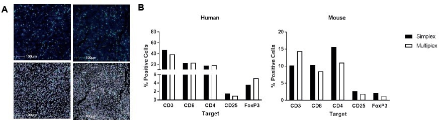
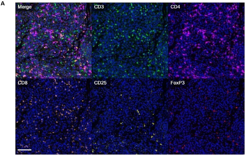
Simplex slides were stained for each individual biomarker in a simplex protocol and compared to a serial section stained with the multiplex IHC. Staining concordance between the multiplex slide and simplex slides was determined by analysis with HALO® after multispectral deconvolution. A) Example images (top) from a simplex slide for target A (left) and the multiplex slide (right) and the corresponding HALO® masks identifying positive cells (bottom). B) Results from the human and mouse Treg panels analysis. The percentage of positive cells from the simplex slide (black bar) is compared to the multiplex slide (white bar). Satisfactory results were obtained for both HISTOPROFILE® -Treg panels. Image Credit: Cerba Research
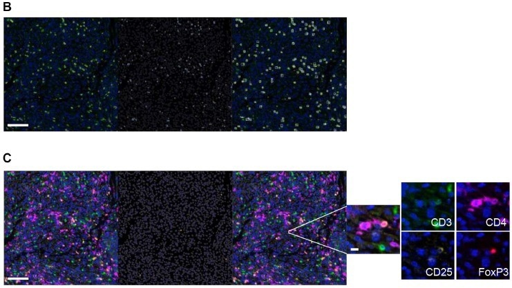
Image Credit: Cerba Research
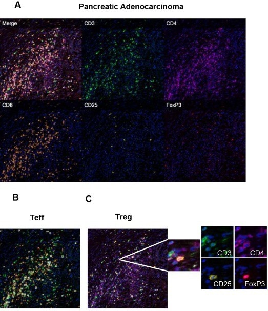
Image Credit: Cerba Research
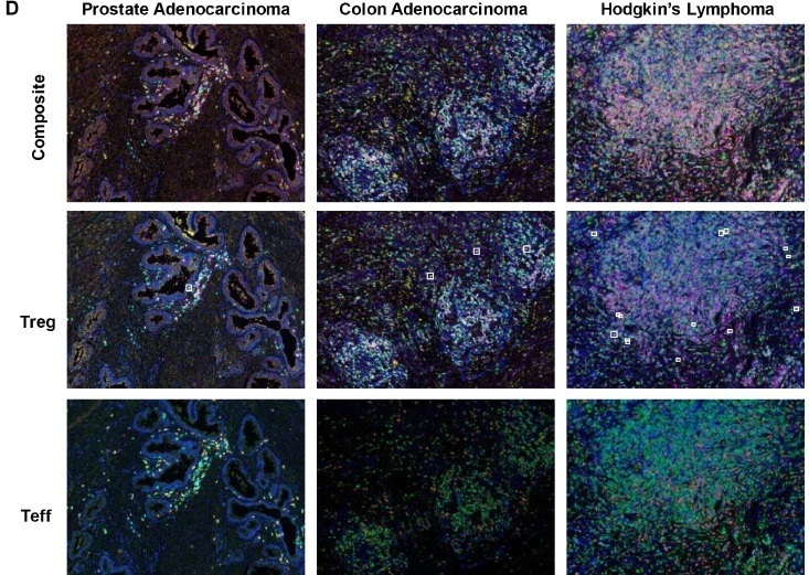
Image Credit: Cerba Research
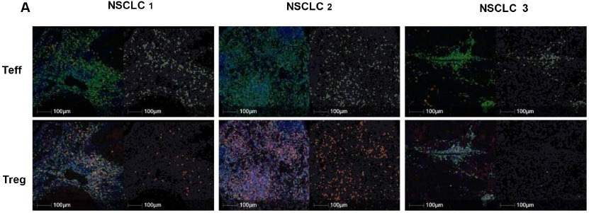
Image Credit: Cerba Research
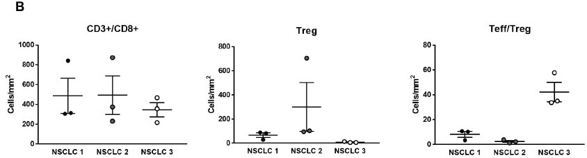
Image Credit: Cerba Research
About Cerba Research
For over 35 years, Cerba Research has been setting the industry standard for exemplary clinical trial conduct. Today, across five continents, with a focus on precision medicine, we are changing the paradigm of the central lab’s role in complex clinical research.
From protocol inception through development and to market, our passionate experts deliver the highest quality specialized and personalized laboratory and diagnostic solutions. Partner with us for the most efficient strategy to actualize your biotech and pharmaceutical products sooner and improve the lives of patients worldwide.
 Brazilian researchers identify cell molecule that drives cancer progression