In this interview, Dr. Michelle Chen, Senior Director of Analytical Sciences at Wyatt Technology, talks to NewsMedical about how to use light scattering techniques to analyze proteins for their multi-attribute quantification (MAQ).
What are the different light scattering techniques used for macromolecule and nanoparticle characterization?
There are three light scattering techniques used for macromolecule and nanoparticle characterization: static, dynamic and electrophoretic.
Static light scattering measures the time-averaged intensity across multiple scattering angles. This method, often called multi-angle light scattering (MALS), provides absolute molecular weight measurements of proteins and other biomolecules in solution, independent of shape. Molecular weight is determined from the total scattered intensity together with molecular concentration, usually measured by UV or differential refractometry (dRI).
MALS can also measure the size and concentration of nanoparticles. Here, the angular dependence of the scattered light yields the size of particles in terms of root-mean-square radius, Rg. For spherical particles, this can be converted to the sphere’s radius, and knowledge of the radius together with total scattered intensity yields particle concentration (particles/mL).
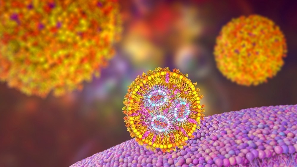
Image Credit: ShutterStock/Kateryna Kon
MALS is used to characterize biological therapeutics like proteins, adeno-associated viruses (AAVs), and lipid nanoparticles (LNPs). The DAWN MALS instrument is typically combined with analytical size-exclusion chromatography, ion-exchange chromatography or field-flow fractionation for initial separation prior to detection, providing detailed analysis. The ultraDAWN MALS instrument, on the other hand, is the process analytical technology (PAT) version of DAWN, used to develop, monitor and control downstream bioprocesses at different scales.
Dynamic light scattering (DLS) measures intensity fluctuations in the scattered light resulting from Brownian motion of the illuminated molecules or particles. By analyzing the time dependence of the fluctuations, one can obtain the translational diffusion coefficient, which is used to calculate the hydrodynamic radius or Rh. Combining DLS with SLS enables estimation of particle concentration, even for heterogeneous sample with multiple size populations.
One popular DLS instrument is the DynaPro Plate Reader, commonly used to screen dozens to hundreds of samples representing different formulation conditions or process fractions. For simpler tasks, the DynaPro NanoStar measures particle size and concentration in microcuvettes with as little as 2 µL of solution.
Electrophoretic light scattering (ELS) is the third light scattering technique, also known as phase analysis light scattering (PALS). It measures the frequency change of the incident laser beam caused by the motion of charged samples in an electric field, from which charge or zeta potential may be calculated.
How does size exclusion chromatography (SEC) differ from field flow fractionation (FFF)?
MALS and DLS detectors are often used with size-exclusion chromatography (SEC) or field-flow fractionation (FFF), also known as asymmetric-flow field-flow fractionation (AF4). Both SEC and FFF provide separation based on a sample’s hydrodynamic volume while the addition of downstream detectors provide in-depth characterization of the sample’s biophysical properties.

Image Credit: ShutterStock/LuckyStep
SEC used a stationary phase consisting of porous beads or resins, with specific pore sizes. Smaller molecules diffuse in and out of the pores, resulting in longer residence time, while larger molecules enter fewer pores and therefore are eluted earlier than smaller molecules.
FFF separates molecules or particles in an open channel, without a stationary phase. During the elution mode, there are two flows: horizontal channel flow and perpendicular cross flow. Smaller particles diffuse against the cross-flow more effectively than larger particles and move further away from the bottom of the channel. Due to the parabolic channel flow profile, the smaller particles are carried by a higher flow velocity relative to the larger particles, so the elution order in FFF is reversed compared to SEC, with small particles eluting first.
Since there is no stationary phase inside the FFF channel, FFF separation is often more appropriate than SEC for samples with dimensions above 30 nanometers, samples that stick to the columns, and/or those that are sensitive to shearing.
What are the main benefits of SEC-MALS for AAV analysis, and how do they compare to other analytical methods? How would you recommend setting up a system and developing fit-for-purpose SEC-MALS methods for AAV analysis?
SEC separation can resolve aggregates and fragments but cannot separate empty and full AAVs. Empty and full viruses will elute in the same peak, as they AAVs have the same hydrodynamic radius. However, with data from the online MALS and concentration detectors, one can calculate empty and full concentrations for the AAV monomer peak and other peaks of interest.
In a single apparatus, SEC-MALS measures molecular weight and concentration of AAV capsids and encapsidated DNA—similar to what one obtains from the combination of ELISA and ddPCR—with an AAV aliquot of just 10 – 100 µL, a short 30-minute runtime and no reagents, saving both time and material while increasing accuracy and precision.
The viral vector analysis module in ASTRA software, provided by Wyatt Technology, calculates several AAV-specific CQAs: the total AAV genomic concentration Vg, the AAV capsid concentration Cp, and Vg/Cp, the full-to-total capsid ratio. ASTRA also calculates the empty AAV concentration, simply Cp minus Vg, and the aggregate content.
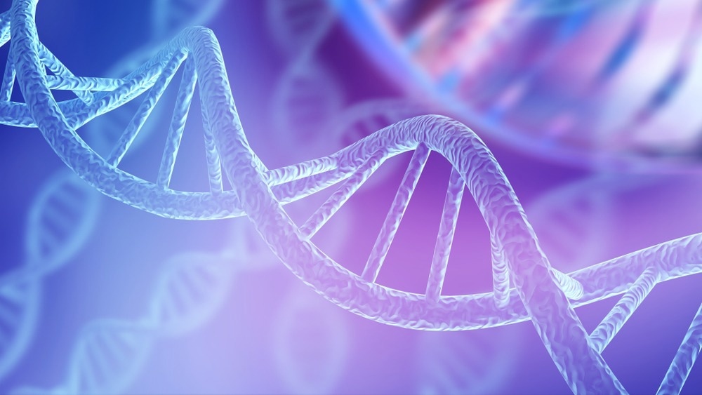
Image Credit: ShutterStock/Billion Photos
The following detectors are recommended for an optimal AAV system for multi-attribute quantification (MAQ): DAWN MALS instrument, Optilab dRI detector and a multi-wavelength UV detector. It is important to have accurate, serotype-specific capsid and DNA UV extinction coefficients. Wyatt Technology provides default extinction coefficients that are reasonably accurate for all AAV serotypes, but more accurate values may be determined with another ASTRA method that utilizes the Optilab and UV detectors.
The extinction coefficient values are then used with MALS-UV-dRI or MALS-UV260-UV280 systems to determine accurate empty and full titers along with Vg/Cp, aggregate species identification and aggregate quantities. Apart from the main CQAs mentioned above (capsid content, capsid titer, genome titer, and aggregation), the same SEC-MALS run can be utilized for extended characterization of protein and DNA molecular weight, capsid integrity, capsid size, and free DNA.
Aggregation content is an important CQA. It is should be noted that since SEC may filter out or disrupt large aggregates, FFF-MALS may be better suited to AAV aggegate analysis.
The SEC condition specified in our Standard Operating Procedure (SOP) Guidance Manual provides good separation of monomers, aggregates and fragments for different AAV serotypes. Wyatt’s AAV SEC-MALS method has been adopted as a platform method for quite a few AAV programs. It is used for multiple serotypes and at different stages of development and commercial production.
What challenges arise in characterizing LNPs compared to proteins and AAVs?
Compared to proteins and AAVs, LNPs are relatively less-well characterized at this stage due to their novelty and the limited availability of modality-specific tools for characterization. The complexity of LNPs arises from their large size and heterogeneity. While protein or AAV samples typically exhibit a smaller number of distinct species, LNP samples often exhibit a continuous distribution in size, molecular weight, and payload.
Several physical attributes of LNPs, listed below, are necessary for characterization and quantification. Using conventional methods, around 10 different assays are required to cover this list.
DLS is often employed to analyze LNP size and polydispersity in support of formulation, process development, and optimization. However, DLS suffers from low resolution compared other light scattering methods like SEC-MALS. Even a small amount of large aggregates can cause a significant change in the average size. Consequently, DLS results are sometimes only qualitative.
The RiboGreen fluorescent assay has been extensively utilized for quantifying RNA payload and encapsulation efficiency. However, this indirect concentration assay can introduce significant experimental errors and user-to-user variation. Furthermore, it does not reveal the variation in RNA payload or dosing with LNP size.
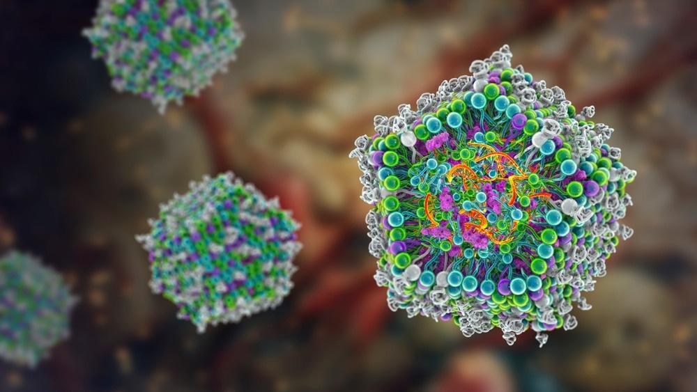
Image Credit: ShutterStock/Kateryna Kon
About Dr Michele Chen
Dr. Michelle Chen is the Sr. Director of Analytical Sciences at Wyatt TechnologyTM, a portfolio of Waters CorporationTM. She obtained her Ph.D. from the Chemical Engineering Department at Yale University, where she focused on developing novel HPLC approaches for high-speed and high-efficiency separations of biopolymers.
Since joining Wyatt Technology, Dr. Chen has incorporated multi-angle light scattering and dynamic light scattering detection with HPLC and field-flow fractionation, for characterizing synthetic and biological polymers, proteins, and nanoparticles.
In recent years she has led the application team at Wyatt to develop novel methods to characterize and quantify emerging bio-nanoparticles including viral vectors, lipid nanoparticles, and extracellular vesicles.
About Wyatt Technology
Wyatt Technology, a division of Waters Corporation, develops instrumentation, software and techniques for the characterization of macromolecules and nanoparticles, in solution, based on light scattering and related technologies. The physical properties determined by Wyatt’s products include absolute molar mass of proteins, polymers and other macromolecules; size and charge (zeta potential); protein-protein and other biomolecular interactions; composition of conjugated proteins and co-polymers; and macromolecular conformation.
Products and services
Wyatt’s product line includes instruments and software for:
- on-line multi-angle light scattering (MALS), used in conjunction with size-exclusion chromatography to quantify absolute molar mass, size, conformation, conjugation and aggregation
- traditional (cuvette-based) and high-throughput (microwell plate-based) dynamic light scattering (DLS) to determine size (radius) and size distributions, protein melting temperature and stability-indicating parameters
- electrophoretic mobility (PALS) to determine molecular charge/zeta potential
- composition-gradient light scattering for label-free analysis of biomolecular interactions
- field-flow fractionation for separation of macromolecules and nanoparticles from 1-1000 nm, used in conjunction with on-line light scattering and other detection technologies to quantify molar mass and size
Wyatt also offers, on a limited basis, sample analysis services utilizing its unique technologies.
_d9995f7d9f4b4ab09c6d14d54eb356e0-80x70.jpg) Wyatt technology announces breakthrough in Field Flow Fractionation (FFF) separation technology
Wyatt technology announces breakthrough in Field Flow Fractionation (FFF) separation technology