A therapeutic monoclonal antibody and its Fab and Fc fragments were recently investigated using differential scanning fluorimetry, temperature-ramped dynamic light scattering, and turbidity measurements.
In an interview with News Medical and Life Sciences, Professor Freiß described how the combination of orthogonal methods allowed a better understanding of the conformational and colloidal stability of monoclonal antibodies.
Why is monoclonal antibody stability so important?
Monoclonal antibodies (mAbs) are an increasingly important aspect of drug development. As the concentration of protein in therapeutic mAb formulations is becoming rather high, stability is more of an issue. The concentration of mAb can be more than 150 grams per litre, and at such high concentrations, special attention is required to stabilize the protein.
Several different aspects must be considered when assessing the stability of mAbs. The three main areas we focus on are chemical stability; conformational stability, which is the persistence of secondary and tertiary structures; and also colloidal stability, which depends on the interactions of the protein molecules in solution.
All three of these are affected by the propensity of the mAb to reversibly interact with other MAb molecules. Such associations can lead to the formation of larger aggregates, which impacts colloidal stability. It is also linked to conformational stability because, upon unfolding, hydrophobic parts of the protein become exposed and are attracted to their counterparts in neighboring molecules .
A pronounced, interactive and attractive reversible self-interaction may be augmented by an additional conformation, stability or unfolding event. If conformational instability occurs, the protein may unfold. However, if there is a repulsive interaction, aggregation may not necessarily occur even if unfolding occurs. Finally, the reversible self-interaction may also affect the extent of the solubility of the mAb, and even cause an increase in viscosity at high concentrations. This can preclude its development as an injectable formulation.
Our research involves analysis of protein-protein interactions by dynamic light scattering and the determination of the effects of temperature on the diffusion coefficient to understand interface-induced mAb aggregation.
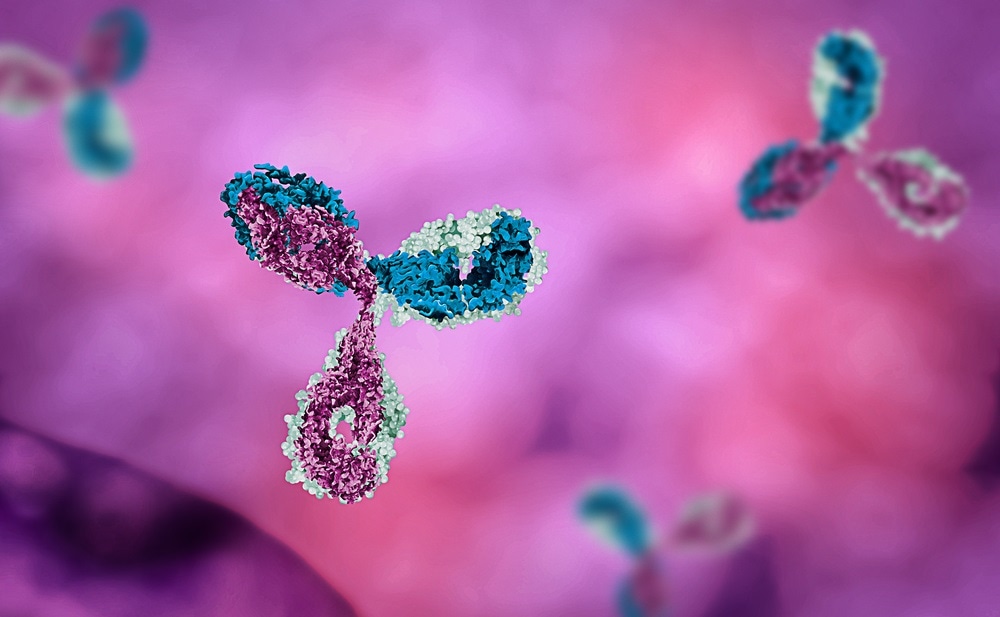
Image Credit:Shutterstock/Mirror-Images
Can you explain the contribution of protein-protein-interactions?
Although protein-protein-interactions occur at low concentrations, they become considerably more relevant at high concentrations. As the concentration of mAb increases, so does the propensity and the probability of interactions.
This is referred to as the molecular crowding effect. There is more excluded volume to which an individual molecule no longer has access, and this restricts its mobility.
The protein-protein interactions may be pairwise or non-pairwise, short-range or long-range. These intermolecular interactions are typically triggered by electrostatic and hydrophobic interactions, which can also cause cluster formation.
How do you study these interactions using DLS?
A non-pairwise interaction can be nicely studied by dynamic light scattering (DLS) as changes in the hydrodynamic diameter can be readily determined using DLS.
The technique was used, along with static light scattering, to evaluate the larger clusters that form by monitoring the apparent hydrodynamic diameter of the antibody. By evaluating the effects of different salts, it was shown that it was the presence of an anion that was important in triggering such a response. Negatively-charged citrate and phosphate anions triggered this cluster formation, whereas the positively-charged histidine buffer did not. The interactions were also found to be pH dependent, with the hydrodynamic diameter increasing from 10 nanometers at pH5 to about 22 nanometers at pH8. With histidine buffer, however, the hydrodynamic diameter hardly changed.
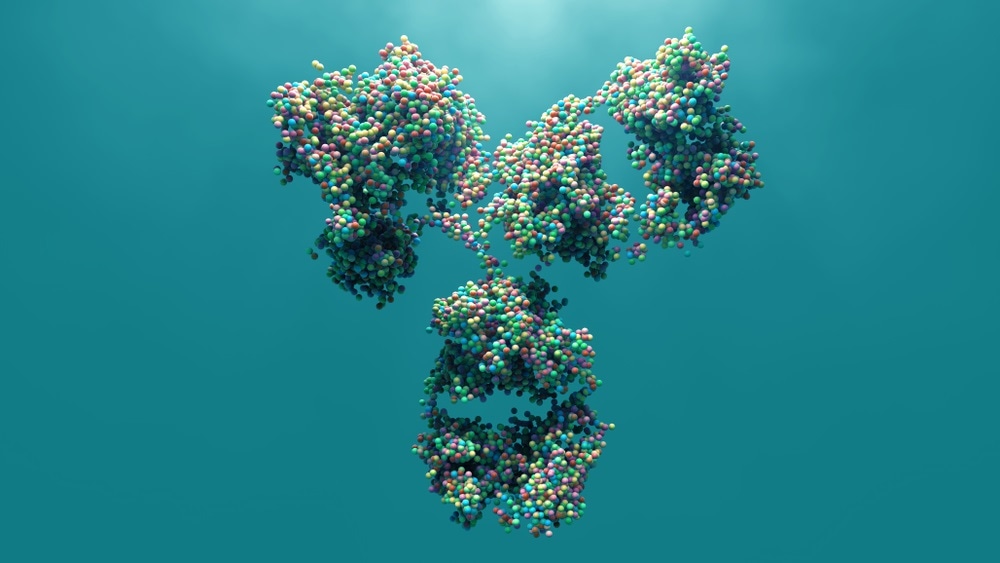
m.jpg)
ImageCredit:Shutterstock/ustas7777777
How does DLS complement other available tools?
Many people start looking at the association of antibodies by using size-exclusion chromatography. Studying 24 different mAb formulations at different pH and sodium chloride concentration, there was no change in UV signal between the different formulations in size exclusion chromatography. Thus, it appears that there is only a monomer present.
Using DLS and asymmetric-flow field-flow fractionation, in addition to size-exclusion chromatography, revealed an interesting mAb behavior. In the absence of salt, pH had a strong effect on the hydrodynamic radius. At pH 7 the radius was almost 20 nanometers, whereas at pH5 it is only six nanometers, which corresponds to the monomer. This cluster formation is suppressed by the presence of 70 or 140 millimolar sodium chloride. Thus mAb association, cannot be seen using size-exclusion chromatography due to the buffer having a higher salt content and the protein being diluted at the detector.
In addition, DLS can be used to quantify the strength of the interaction by facilitating kD analysis. Also, only small volumes are needed - we measure down to volumes as low as five microliters - and this facilitates temperature ramps.
What can we learn from determining kD with DLS?
We can derive kD from the slope of the apparent diffusion coefficient as a function of the protein concentration. A positive slope indicates net repulsive interactions, whereas a negative slope points towards net attractive interactions. The kD obtained in this way can then be compared to the standard A2 values, using corrections for the molecular weight, the friction coefficient, and the specific volume.
We have looked at various different antibodies in different studies and tried to correlate their kD with their A2 values. Using histidine buffer, citrate buffer and acetate buffer, at different pH and in the presence of sodium chloride or arginine hydrochloride, we showed that an increase in ionic strength leads to an increase in kD. This, however, is pH-dependent. At pH 5 there is no influence of ionic strength observable, and the kD values are higher than at pH 6. On reaching pH 4 the interactions become repulsive. This is correlated to the charge on the protein molecule, meaning that at the lower pH it's more positively charged, and we have stronger repulsive interactions.
In many cases, kD is also correlated to viscosity. There are cases though where there's no correlation between kD and viscosity, especially if the viscosity gets really high.
kD can be used as a parameter to study antibodies and to understand their behavior at high concentration and how the viscosity increases, although the studies are conducted at low concentrations.
How is kD affected by temperature?
It is important to assess the effects of temperature on a protein’s propensity to unfold and aggregate. Using DLS we can determine the temperature at which the protein starts to unfold, and aggregation occurs—the temperature of aggregation (Tagg). When the kD is higher, the temperature at which aggregation starts is higher because we are starting with more repulsive interactions. MAbs with a lower kD start to unfold at a lower temperature and their propensity to form aggregates is higher. So, a lower kD means a lower Tagg. Such studies help us select the most stable formulation of a particular mAb by identifying which conditions are destabilizing and which are stabilizing.
DLS can become a complimentary method for temperature-dependent studies, to support typical temperature ramp methods like differential scanning calorimetry (DSC). It can also be supported by temperature ramped UV-VIS, fluorescence, or infrared. It can distinguish how effects differ between different sections of a molecule. For example, hydrophobic and hydrophilic areas will unfold in different conditions. This is especially important for mAbs as these comprise distinct Fab and Fc parts.
Investigating changes to the Fab and the Fc part of the antibodies separately, allows you to learn more about the molecule. With cetuximab, a detailed DSC analysis showed that the Fab part starts to aggregate as soon as it starts to unfold, whereas the Fc part shows two different transitions. The first part of unfolding already starts at temperatures well below 70 degrees C, but does not cause aggregation. Aggregation is only triggered at about 75 degrees C, when the second unfolding starts to occur.
By adding DLS analysis of cetuximab, we could see that the hydrodynamic diameter increases substantially as the temperature rises. The hydrodynamic radius and kD remained unchanged up to about 60 degrees C, unfolding then starts to occur, and this leads to a substantial change in the diffusion coefficient. Meaning that we all of a sudden have larger species, and a considerable decrease in kD. This means that substantial interactions between the molecules are occurring before we see noticeable precipitation. Similar behavior was observed when the Fc part was studied separately.
The DSC analysis showed that the Fab part started to aggregate at the beginning of any unfolding event. However, a decrease in kD observed with DLS as the unfolding began was not accompanied by substantial precipitation. The aggregation occurs at temperatures higher than 80 degrees C.
The combination of studies revealed that the conformational stability of monoclonal antibodies is determined by the net charge due to the effects on electrostatic attractive and repulsive interactions. Conformational stability is thus greatest at higher pH, without or with salt, and also at lower pH with salt. Temperature-induced unfolding is triggered aggregation by exposing hydrophobic patches that mediate a reduction of kD.
Does mechanical stress also affect kD?
As with thermal stress, mechanical stress, such as shaking and stirring, can cause large aggregates to form. Similarly, the susceptibility to mechanically-induced aggregation is also determined by the formulation.
There's a hypothesis that these protein-protein-interactions are important for the formation of these aggregates upon mechanical stress. The study by He et al. showed that with a higher kD, the particle formation upon mechanic stress and formulation of one mAb was substantially reduced, whereas, if kD was equal to zero, there was substantial particle formation. There's not, let's say, a real line of correlation into that, but it really tells you that the repulsive interaction may lead to the fact that, upon mechanics stress, we have less aggregation. We studied that.
We conducted infrared (IR) spectroscopy at the mAb interface and this showed that there's a film formation at the interface. We evaluated the IR spectra, and they're very similar to what we see in solution. We cannot really tell whether the protein is unfolded or not. We don't see a pH effect on the IR spectra.
Brewster Angle Microscopy showed clusters of protein within the film. You could even compare it to the Arctic sea where you may have some ice shields and floats on the different ice shield, so it's all overlapping within a protein film.
Compression of the film results in some aggregate or cluster formation. When we shake up the mAb, increasing pH results in stronger particle formation. kD decreases with increasing pH. We, therefore, have this correlation between particle formation and the kD value that we can measure to determine the propensity for aggregate formation at the interface.
We also used DLS to study mAb conjugates and found that introducing an additional hydrophobic moiety makes the attractive forces more pronounced and kD values decrease. The kD analysis thus helps us to understand and predict the behavior of conjugates as well.
ImageCredit:Shutterstock/Design_Cells
Can you summarise the key findings?
In summary, the DLS measurements and the A2 measurements in mAb formulations provide us with information on protein-protein-interactions, in particular, at storage conditions. The A2 can actually be a missing link in temperature- and interface- induced mAb aggregation.
Repulsive net charges at low pH increase the colloidal stability, although a reduction of the conformational stability was also observed. Aggregation was driven by hydrophobic interactions. Coupling of differently charged hydrophobic payloads affected colloidal stability mostly based on the charge effects.
About Professor Wolfgang Frieß
Professor Wolfgang Frieß is a professor of pharmaceutics and biopharmaceutics at Ludwig Maximilians Universitaet Munich, Germany. A key element of his research is investigating the link between the thermal unfolding and aggregation of proteins in order to help to minimize conformational and colloidal instabilities in therapeutic proteins formulations.
-2.jpg)
 Control and Monitor Nanoparticle, Biopharmaceutical and Polymer Product Process