In this interview, News Medical speaks with Professor Govind Srimathveeravalli, Associate Professor of Mechanical and Industrial Engineering at the University of Massachusetts Amherst, about a new approach to treating bladder cancer that uses electrical energy to target cells lining the bladder, aiming to reduce recurrence and help more patients avoid radical surgery.
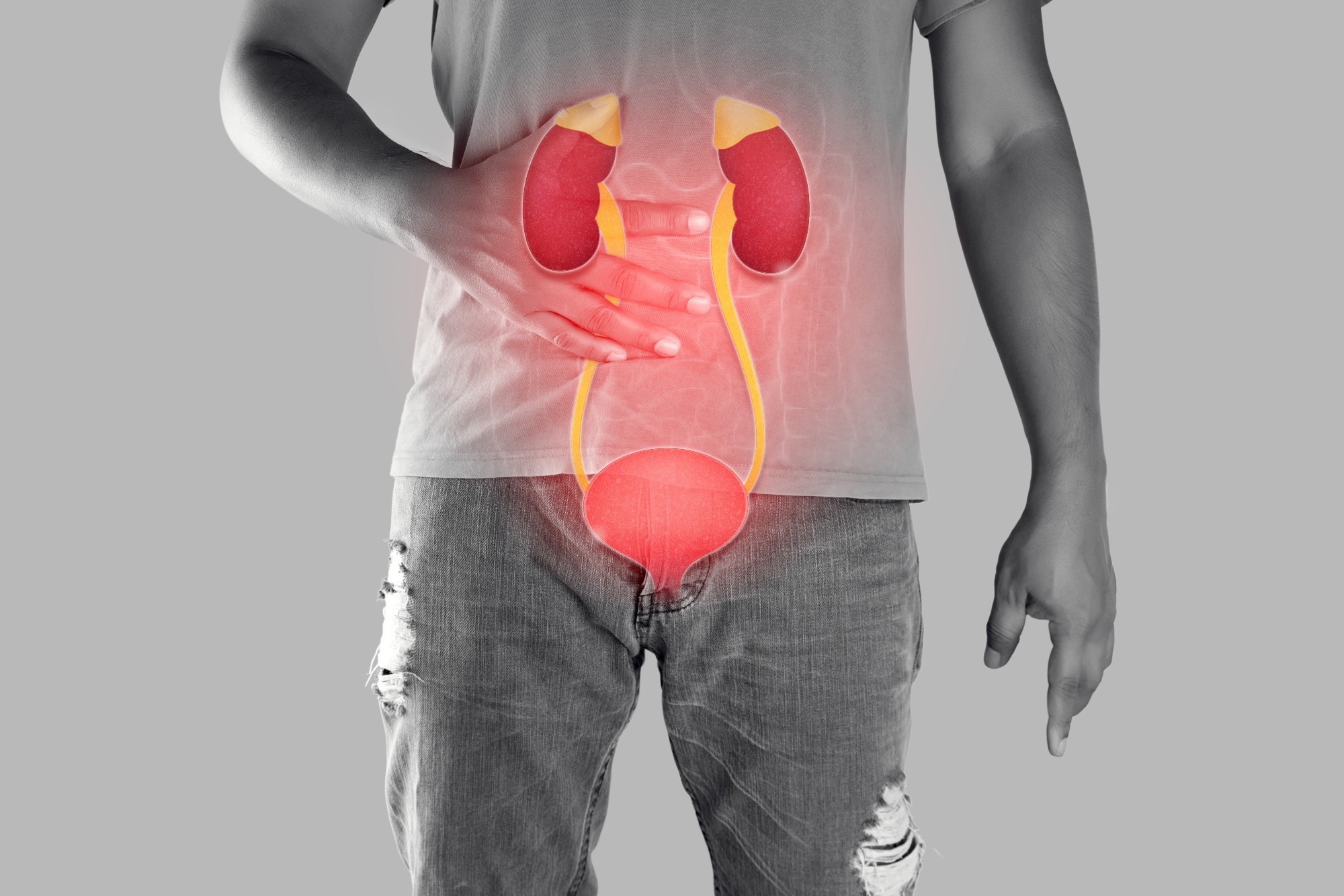 Image credit: Emily frost/Shutterstock.com
Can you please introduce yourself and your role at the University of Massachusetts Amherst?
I am currently an Associate Professor at the University of Massachusetts Amherst, where my lab develops medical devices for minimally invasive, non-surgical therapies. I joined UMass Amherst in 2019, and before that I spent about eight years at Memorial Sloan Kettering Cancer Center in New York City.
A major focus throughout my work has been developing medical devices that allow us to treat cancer without conventional surgery. One technology I have worked with extensively is irreversible electroporation, or IRE, which is more recently also referred to as pulsed field ablation.
IRE delivers a very small amount of electrical energy into tissue. It is not radiation. We use electricity to kill cells without overtly damaging the underlying structure of the organ or tissue where those cells are located. This is particularly useful when treating areas close to sensitive structures such as the bladder, urethra, ureter, or bile ducts.
My lab has been pursuing this area for almost 15 years. We are particularly interested in tumors that develop within tubular organs because these can be some of the most challenging cancers to treat surgically. Surgery may cure the patient, but removing part or all of an organ can have significant consequences for their quality of life.
Why is bladder cancer recurrence such a persistent challenge for patients and clinicians?
For many patients, bladder cancer can be associated with repeated environmental exposure to certain toxins. This could include smoking or occupational exposure to chemicals, for example.
One challenge is the field cancerization effect. Bladder cancer develops from a thin layer of cells lining the bladder called the urothelium. These cells perform an important barrier function, keeping urine on one side and the rest of the body on the other.
In some patients, it is not simply one isolated group of cells that has become problematic. A much larger area of the bladder lining may have been affected. Some cells have already become malignant, while others may have the potential to become malignant in the future.
A procedure such as transurethral resection of bladder tumor (TURBT) can remove the tumors that a clinician can see. However, microscopic disease or abnormal cells may still be scattered elsewhere in the bladder. That is why additional treatments such as chemotherapy or immunotherapy are used after TURBT. The aim is to clear the remaining microscopic disease.
The problem is that recurrence remains very common. About 50% of these patients experience recurrence within two years, and if you follow a relatively young patient long enough, they may have multiple recurrences. Eventually, the disease can become more aggressive, and the options become increasingly limited.
You have compared bladder cancer recurrence to a game of "whack-a-mole." How does that analogy explain the problem your team is trying to solve?
The idea is that diseased cells can hide among healthy cells in the bladder lining. Current drugs are essentially trying to find and kill those hidden cells. If the treatment doesn't eliminate everything, the disease can come back.
You can then try another drug and perhaps another treatment, but eventually, if those approaches fail, you may reach the point where the bladder has to be removed.
Our approach is different because we are not trying to discriminate between an individual healthy cell and an individual diseased cell within this very thin layer. Instead, we are asking whether we can treat the layer itself.
Think about exfoliating skin. You remove an outer layer and fresh cells regenerate from underneath. We are asking whether we can do something conceptually similar with the urothelium.
Using electrical energy, we want to remove or kill the problematic cellular layer while preserving the underlying structural tissue. If the underlying tissue is healthy, we hypothesize it can regenerate into a healthy bladder lining.
That is what we are betting on, and that is also what makes this an interesting engineering challenge. The layer we want to target is extremely thin, so we need to deliver the treatment with great precision.
How does the irreversible electroporation technology work, and what could treatment look like for a bladder cancer patient?
The technology uses electrical energy to kill cells without overtly damaging the underlying tissue structure.
In the clinic, IRE is already used in other settings. Physicians can insert thin needles into a tumor, deliver electrical pulses, and kill the targeted cells. The body can then absorb what remains.
For bladder cancer, however, we are developing a catheter-based device. The idea is that a physician would introduce the device into the bladder, treat the bladder lining using electrical pulses, and then remove the device. This is a procedure, not a catheter that remains inside the patient.
Our treatment itself is extremely rapid. In the laboratory, the treatment can be performed in approximately 10 minutes. Of course, translating that into human patients requires further development and validation, but ultimately we envision a relatively short procedure performed under appropriate anesthesia.
We believe this could potentially reduce the risk of resistant disease and recurrence, but we do not know that yet. Establishing whether that is actually the case is part of the research we now need to conduct.
Discover How Electrical Pulses Could Help Prevent Bladder Cancer Recurrence: Click Here to Download your Free PDF
Where would this treatment fit into the current bladder cancer treatment pathway?
This is an important distinction because we are not proposing this technology as an alternative for every patient with bladder cancer. Our intervention would occur earlier in the treatment pathway.
For example, a patient could undergo TURBT, where the clinician removes all the tumors that are visible during cystoscopy. After that, patients may receive intravesical chemotherapy or Bacillus Calmette-Guérin (BCG) immunotherapy to address microscopic disease remaining within the bladder.
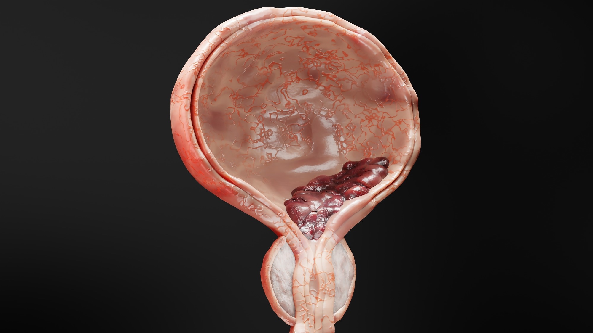 3D illustration of a malignant tumor within the bladder. The research aims to intervene after visible tumors have been removed, targeting residual disease in the bladder lining before it can recur or progress. Image Credit: ALIOUI Mohammed Elamine7/Shutterstock.com
We are asking whether our treatment could be used at that stage instead. After removing the visible tumors, we would use electrical treatment to non-specifically clear the problematic cellular layer throughout the bladder.
The hope is that this could prevent or reduce recurrence and, importantly, prevent progression to muscle-invasive bladder cancer.
Once the cancer has become muscle-invasive and is growing into the body rather than simply into the bladder cavity, it may be too late for the technology we are developing. At that stage, surgery can be necessary.
Our goal is therefore to intervene earlier so that fewer patients ever reach the point where radical surgery is required.
Why is avoiding cystectomy such an important goal beyond simply treating the cancer itself?
When somebody is diagnosed with cancer, their immediate priority is understandably to get rid of the disease. But successful cancer treatment can come at a cost, and patients do not necessarily experience all of that cost immediately. Some consequences appear later.
Removing the bladder is a major operation. To create a urinary diversion, surgeons may use a section of the patient's intestine to form a conduit or pouch. That intestinal tissue still has intestinal functions. It contains bacteria, produces mucus, and absorbs electrolytes, all of which can create additional complications.
We have actually been investigating another application of IRE that addresses this problem.
Immediately before the surgeon uses the intestinal segment to create the urinary diversion, we can treat that tissue using IRE to remove the intestinal cells while preserving its underlying structure.
The idea is that once this tissue is connected to the urinary tract, urinary tract cells can populate that structure. Potentially, the tissue could then behave more like part of the urinary system rather than continuing to behave like intestine.
We have been working on that research for around five years. More broadly, our lab is developing multiple technologies around bladder cancer, both to prevent patients from requiring radical surgery and to reduce complications when surgery cannot be avoided.
How important is your collaboration with clinicians such as Dr. Jonathan Coleman at Memorial Sloan Kettering Cancer Center?
It is extremely important because my lab is focused on translational research.
Curiosity-driven research involves scientists identifying an interesting biological or technical question and investigating it. Our approach starts somewhere different. We start with the clinical problem.
In this case, bladder cancer recurrence is the problem. Clinicians are using different drugs and therapies, yet recurrence remains a significant challenge, and some patients ultimately require bladder removal.
We start with that problem and ask: what laboratory tools might address it?
Clinical collaborators help determine what evidence matters, which experiments to perform, and how to design the technology so that, if we demonstrate safety and effectiveness and show it can scale to human anatomy, we are positioned to move toward clinical use.
Even then, developing the technology is only half the battle.
A medical device must also overcome regulatory, commercial, and reimbursement challenges. Physicians need to be convinced that a new treatment offers a meaningful advantage over something they already use and trust.
That is why clinical partnerships are critical throughout the translational process.
You now have five years of grant-supported research ahead. What needs to happen before this technology could reach patients?
A five-year grant might sound like the beginning of the research, but grants are often more of an inflection point.
Reaching this stage means we already have data suggesting we have a potential solution and that it could work. The grant allows us to accelerate the additional experiments needed to investigate that possibility.
If we gather the evidence we need and the results show the approach is safe and effective, the next stage would involve finding a commercial partner willing to invest in translating the technology.
The prototypes we build in an academic laboratory are not the devices that ultimately go into patients. An industry partner would be needed to manufacture the technology at the right scale and to the required standards.
We would also need to engage with the regulatory authorities and establish exactly what evidence is required before the device could be approved for human use.
Scientific evidence is essential, but taking a medical device to patients requires a different body of evidence, along with regulatory, manufacturing, and commercial expertise.
Could the same technology eventually be used beyond bladder cancer?
Potentially, yes. The technology we are developing could have implications for certain gastrointestinal conditions and cancers.
One example is Barrett's esophagus, which can develop in people with prolonged acid reflux. Repeated exposure to acid changes the cells lining the esophagus and can increase the risk of esophageal cancer.
In theory, a similar technology and device concept could treat the affected cellular layer.
Another potential area is colorectal disease. The same kind of field cancerization effect that contributes to repeated bladder cancer recurrence can also result in larger fields of polyps in the colon that may later become malignant. Currently, clinicians can remove individual polyps, but there is no straightforward way to treat the wider tissue bed containing potentially diseased cells.
There may therefore be several applications for this concept.
However, regulatory approval for one indication does not automatically translate to another. Even if we demonstrated that the technology was safe and effective for bladder cancer, using the same device for another disease would require us to go back and establish the evidence required for that particular application.
What would you most like clinicians, researchers and medical technology companies to take away from this research?
One of the main reasons I wanted to discuss this research is that we are always looking for partners.
I am interested in clinicians who may look at this technology and recognize an application we haven't considered. There will inevitably be blind spots because the applications we currently pursue are shaped by our own knowledge and experience. Someone else may understand a clinical problem where this technology could be an excellent fit.
We are also interested in industry partners who see the translational potential and want to discuss how the technology might be developed further.
Scientific publications are incredibly important because of their rigor, but relatively few people read them in depth. Communicating the research more broadly can help us reach clinicians, engineers, and companies with different perspectives on where the technology could make a difference.
Ultimately, the goal is not simply to develop something interesting in the laboratory. We want to translate these technologies into solutions that can reach patients.
About Professor Govind Srimathveeravalli
Professor Govind Srimathveeravalli is an Associate Professor of Mechanical and Industrial Engineering at the University of Massachusetts Amherst, where he is also an adjunct faculty member in Biomedical Engineering and Graduate Program Director in Mechanical Engineering. He directs the Center for Personalized Health Monitoring within the university's Institute for Applied Life Sciences.
His research focuses on the interaction between non-ionizing energy and biological tissue and on translating those principles into novel medical devices. His work encompasses minimally invasive cancer therapy, tumor ablation, drug delivery, and tissue engineering, with a particular focus on developing technologies that deliver electrical energy to difficult-to-access locations within the body. Before joining UMass Amherst in 2019, he spent about eight years at Memorial Sloan Kettering Cancer Center.
Professor Srimathveeravalli received his Bachelor of Engineering degree from the University of Madras in Chennai, India, before completing both his MS and PhD at the State University of New York at Buffalo. His research has received support from organizations including the National Institutes of Health, the National Science Foundation, and the Department of Defense. In 2026, he was named a Senior Member of the National Academy of Inventors, recognizing his work translating patented medical technologies toward real-world applications.
 UCLA researcher wins the 2026 Albany Prize for revolutionary cancer therapy discoveries