Sponsored Content by RefeynReviewed by Olivia FrostJul 18 2024
Oligomerization has the potential to be a key factor in protein function, so properly understanding the function of a protein necessitates the quantification of its oligomerization state.
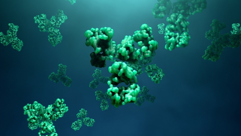
Image Credit: Design_Cells
Mass photometry offers a means of measuring a sample’s mass distribution in native conditions and at a single-molecule level, with this technique even being sensitive enough to detect rare species.
Automated mass photometry builds on these capabilities with easy and consistent sample dilution and manipulation, enabling thorough assays of oligomerization behavior.
Oligomerization - best understood as self-assembly into specific quaternary structures - is one of the core processes for many proteins’ functions. Proteins that function as monomers are actually in a minority, according to current databases.1,2
Oligomer formation has a number of relevant physiological roles. For example, gene expression regulation typically relies on homo-oligomeric DNA-binding proteins. It is also possible for enzyme activity to be regulated via allosteric interactions between subunits or through the formation of active sites at the subunit interface.
Membrane-associated proteins (such as receptors and channels) often require oligomerization to provide transport across membranes or to enable cell-to-cell adhesion.2,3 Altered oligomeric states are also a factor in disease development in some instances, making these especially relevant in the context of translational research.4
By evaluating and identifying conditions that determine a given protein’s oligomeric state, it is possible to then develop mechanisms for interfering with or stabilizing this process. By doing so, it is possible to explore new and potentially valuable avenues of therapeutic intervention.
Mass photometry and protein oligomerization
It can be especially challenging to properly capture and characterize protein oligomerization if the concentrations of species in the sample are very low.
Mass photometry is one of the most suitable techniques available for the assessment of oligomerization states and dynamics. This powerful technique works by measuring the amount of interference between light reflected by a glass surface and light scattered by a molecule in contact with the glass.
The magnitude of the interference scales linearly with molecular mass. Notably, this interference can be measured without labels, using a wide range of buffers, and with minimal sample amounts.
Mass photometry affords users high-resolution distributions of a sample’s molecular mass directly in solution while maintaining single-molecule sensitivity. The information about the mass distribution of a sample can then be used to infer oligomerization states.
Mass photometry even offers sufficient sensitivity to detect rare species comprising less than 1 % of the main sample population.
Automation of this analytical process via Refeyn’s Auto platform offers a key advantage that complements mass photometry’s inherent strengths. The Auto robotics unit is fully compatible with Refeyn’s OneMP and TwoMP mass photometry instruments, allowing users to automate their mass photometry processes easily.
This combination of sensitivity and efficiency makes this system ideally suited to the rapid screening and analysis of oligomeric states under a range of conditions.
Mass photometry can characterize oligomerization behavior
The experiment series discussed here was performed by GSK using the TwoMP Auto. The company’s team undertook a preliminary study in order to characterize the oligomerization dynamics of a proprietary protein of interest.
To achieve this, the team assessed the protein’s oligomeric status at a number of different concentrations. In the buffer alone, it was noted that the target protein primarily formed monomers at lower concentrations.
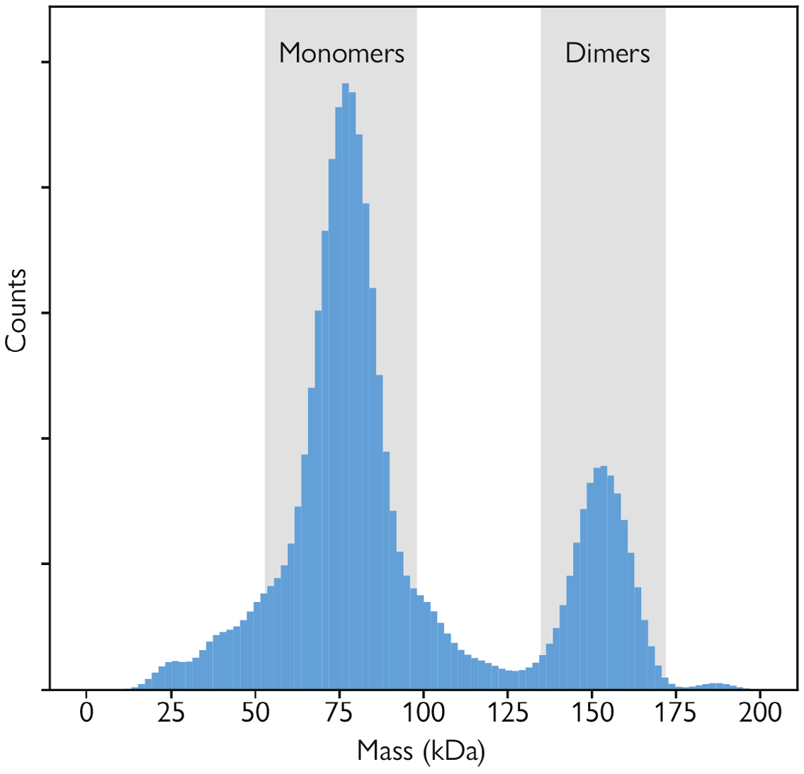
Figure 1. Measuring oligomerization with mass photometry. Mass distribution of a sample of the target protein measured at a concentration of 6.3 nM. The peak containing the main population (around 80 kDa) corresponds to counted monomers, while the secondary peak (around 160 kDa) corresponds to dimers. Image Credit: Refeyn Ltd.
It was also observed that as the protein’s concentration increased, homodimers became apparent in the mass distribution histogram. These can be seen as a secondary peak at twice the molecular mass of the monomer peak. It was also noted that both peaks were visible at a protein concentration of 6.3 nM (Figure 1).
Mass photometry helps study oligomerization effectors
The impact of varying calcium concentrations was evaluated in this experimental series, and the addition of a candidate inhibitor molecule that had been hypothesized to prevent oligomerization was tested.
A series of measurements were undertaken to increase protein concentrations following the addition of the candidate inhibitor. Both sets of measurements were streamlined via automation, and these were used to calculate the dissociation constants of the homodimers both with and without the presence of the inhibitor (Figure 2).
Results of these experiments implied that the candidate inhibitor impacted protein oligomerization, leading to notably reduced dimer abundance in the presence of the inhibitor.
This change was measured by calculating the dissociation constant (KD) of protein samples without inhibitor (KD= 3.55 ± 1.2 nM) and also with inhibitor (KD= 9.1 ± 1.1 nM). The effect was found to be minimal, suggesting there may be a different inhibition mechanism in this case.
It was also observed that oligomerization was impacted by the presence of calcium. Tetramers were found to be promoted by the addition of CaCl2, appearing as a peak on the mass distribution histogram at four times the monomeric species’ molecular mass.
The addition of the calcium chelator EDTA was found to reverse the formation of tetramers, with the histogram reflecting this as the absence of the third peak (Figure 3).
The team further explored the impact of calcium on target protein oligomerization by acquiring repeated mass photometry measurements at increasing protein concentrations while maintaining constant calcium concentrations.
The presence of calcium resulted in a gradual increase in tetramers as protein concentration increased. This also coincided with a reduction in monomers (Figure 4) while the proportion of dimers remained constant. This was in contrast to the behavior noted in the absence of calcium (Figure 2).
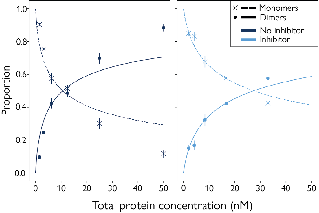
Figure 2. Characterization of protein oligomerization with and without an inhibitor. Relative proportions of monomers and dimers in the sample, as a function of target protein concentration. Left plot (dark blue) shows measurements in simple buffer, while right plot (mid blue) shows measurements in the presence of an oligomerization inhibitor. Concentrations measured were : 1.55, 3.15, 6.25, 12.5, 25.0 and 50.0 nM for the samples without inhibitor, and 2.1, 4.2, 8.4, 16.7 and 33 nM for the samples with inhibitor. Image Credit: Refeyn Ltd.
Automated mass photometry streamlines oligomerization research
The TwoMP Auto’s automated sample manipulations mitigate the variability commonly linked to manual operation between operators and across different experiments.
Reproducibility testing with the TwoMP Auto highlighted less than 1 % variabilities in the measured mass and relative proportion of detected species. This quality was especially beneficial for GSK’s preliminary study and its characterization of the investigated candidate inhibitor.
The TwoMP Auto’s ‘in-plate’ dilution feature also proved a crucial feature in this series of experiments due to its ability to store the protein sample in each well at a higher concentration before diluting this immediately before measurement takes place.
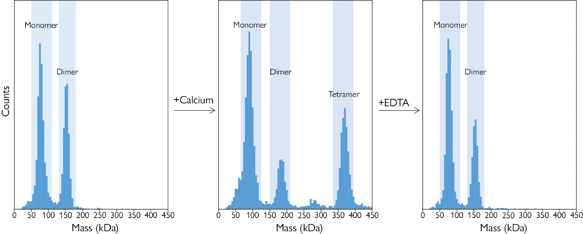
Figure 3. Reversal of tetramer formation. A: A mass histogram of a protein-only sample, with monomers and dimers present. B: Calcium chloride (present in excess) induced the formation of tetramers and reduced the number of dimers. C: Adding a saturating concentration of EDTA reversed this effect, resulting in the disappearance of the tetrameric species. Image Credit: Refeyn Ltd.
In-plate dilution also helps to minimize any potential adsorption to the well surface, a significant issue at low protein concentrations.
This product feature minimizes the need to manually dilute samples to the required concentration range for optimal mass photometry measurements. The system also allows users to run several sets of conditions in each run, reducing the required overall screening time.
Using Refeyn’s DiscoverMP software, all files generated from each run can be analyzed as a single batch, further simplifying data reporting and streamlining data analysis.
In summary, mass photometry represents a robust and valuable technique for the accurate characterization of oligomer formation. It is also useful in better explaining factors involved in inhibiting and promoting this crucial biological process.
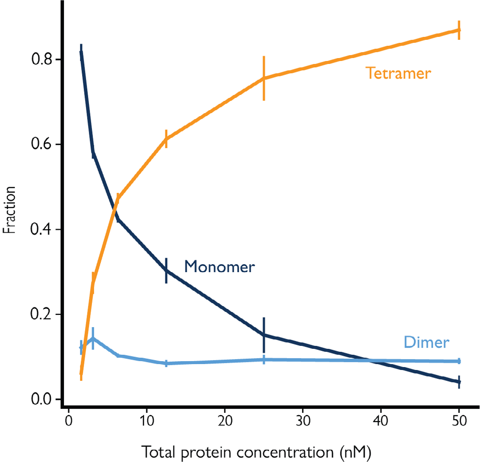
Figure 4. Dilution series showing protein oligomerization under constant CaCl2 concentration. In the presence of 20 mM CaCl2, increasing the target protein concentration resulted in an increase in tetramer formation, while the proportion of dimers in the sample remained small. Image Credit: Refeyn Ltd.
This process can be further streamlined via automation, with improved efficiency in steps from sample preparation through to data capture. The use of automation not only saves the operator time but also promotes the rapid acquisition of reproducible results and a more cost-effective investigation of oligomerization behaviors.
In summary, studying oligomerization using mass photometry offers a range of benefits, including:
- Minimal sample preparation requirements
- The acquisition of accurate results within minutes
- Compatibility with most buffers
- No requirement for the use of labels
- Sensitivity to changes in oligomerization status, even with minimal sample quantities
- Potential to detect even rare oligomeric species
- Rapid, unattended measurement of up to 24 samples via automation
- Suitability for screening and titration, studies of protein interaction and investigation into effector dynamics
References and further reading
- Danielli et al., Sci Rep 2020
- Hashimoto and Pachenko, PNAS, 2010
- Marianayagam et al., Trends Biochem Sci 2004
- Sato et al. J Biol Chem 2018
Acknowledgments
Produced from materials originally authored by Refeyn Ltd.
About Refeyn Ltd.
Refeyn are the innovators behind mass photometry, a novel biotechnology that allows users to characterise the composition, structure and dynamics of single molecules in their native environment. We are producing a disruptive generation of analytical instruments that open up new possibilities for research into biomolecular functions.
Spun out of Oxford University in 2018 by an experienced team of scientific professionals, Refeyn aims to transform bioanalytics for scientists, academic researchers, and biopharma companies around the world.
Sponsored Content Policy: News-Medical.net publishes articles and related content that may be derived from sources where we have existing commercial relationships, provided such content adds value to the core editorial ethos of News-Medical.Net which is to educate and inform site visitors interested in medical research, science, medical devices and treatments.