Drugs and fibers are among the materials collected in forensic and criminal laboratories. Some of these samples can be quite small, and light microscopes are frequently employed to examine evidence gathered at the crime scene.
An optical microscope can help investigators see the evidence more clearly, particularly at the microscopic level. However, in some cases, more information is required to prove beyond a reasonable doubt whether a person is guilty or innocent.
As a result, a reliable and adaptable analytical technique is required to offer visual and chemical information.
Fourier transform infrared spectroscopy (FTIR) is a macroscopically useful technique for forensic scientists. FTIR microspectroscopy expands the application of standard FTIR by enabling rapid, nondestructive investigation of samples as small as 10 microns.
The Thermo Scientific™ Nicolet™ iN10 Infrared Microscope combines an optical microscope and an integrated FTIR. The Nicolet iN10 analytical equipment allows forensic scientists to visually and chemically evaluate illicit pills, hair, fibers, inks, and paints.
The Nicolet iN10 is a powerful, small FTIR microscope thanks to its integrated architecture, which eliminates the need for an external spectrometer.
Evidence is an important aspect of any court case. For the first time, the unique ability to check microscope performance using software gives the investigator and the jury confidence that the data is reliable.
The Nicolet iN10 may be used without liquid nitrogen, allowing the lab to quickly evaluate evidence in any location. The Thermo Scientific™ OMNIC™ Picta™ Software simplifies and speeds up microscopy operations, even for inexperienced users. Powerful wizards assist the user with reflection, transmission, and ATR analysis.
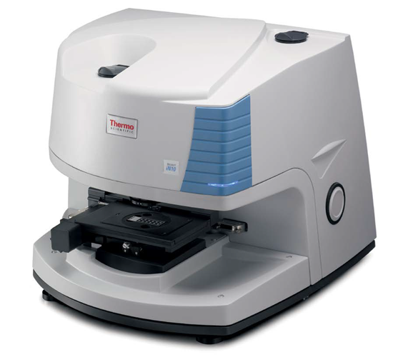
Image Credit: Thermo Fisher Scientific - Vibrational Spectroscopy
Ink on paper
Counterfeiting is one of the earliest known illegal activities. Technological improvements have enabled criminals to create counterfeit notes without using highly sophisticated offset printing procedures. An inexperienced person with access to a photographic copier or scanner can create high-quality counterfeit cash.
However, the paper and ink used in the printing process have different properties that can be used to identify counterfeit notes and even track their origins. Ink is typically evaluated using elemental analysis, X-rays, and mass spectroscopy. These methods provide thorough characterization but are damaging and time-consuming.
Because of the high infrared absorbance from cellulose between 1200-950 cm-1, infrared spectroscopy has not been properly employed in the identification of ink and pollutants on paper.
The rapid and non-destructive nature of infrared imaging and Attenuate Total Reflectance (ATR) FTIR microscopy provides a significant benefit for assessing criminal evidence. Thermo Scientific Nicolet iN10 Microscope can now be used to analyze fake documents.
Analyzing suspect inks can disclose the type of ink and how it was applied to the paper. Visual inspection often distinguishes between ink applied using photostatic or inkjet processes and offset printing techniques. However, with modern printing technology, this is getting increasingly challenging.
FTIR microscopy enables rapid chemical imaging of both ink and paper materials. This yields unambiguous data that may be directly compared to actual documents.
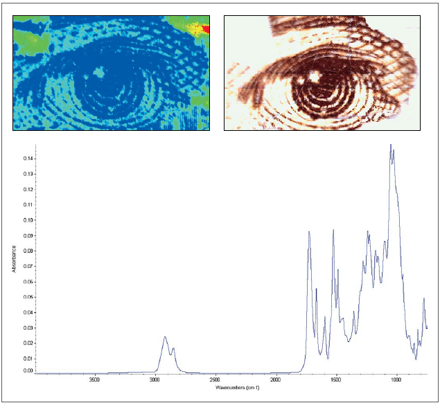
Figure 1. (Upper left) Chemical image of Andrew Jackson’s eye on $20 U.S. currency (Upper right) Video mosaic of Jackson’s eye (Lower) Black ink spectrum collected by Tip ATR. Chemical imaging highlights the distribution, while ATR analysis provides detailed spectral information of the ink. Image Credit: Thermo Fisher Scientific - Vibrational Spectroscopy
Figure 1 depicts a chemical picture of a US twenty-dollar bill. The black ink is chemically distinct from the paper and the surrounding background ink. The chemical and visual pictures can be contrasted, demonstrating the remarkable resolution of infrared microscopic data.
Once the desired information has been discovered using rapid chemical imaging, ATR analysis can offer detailed spectrum data with minimal interference from the cellulose contribution.
Fiber and hair analysis
Many fibers are often discovered at crime scenes and can provide useful or even critical information.
Forensic scientists, for example, are trained to recognize and associate physical hair traits and looks with specific ethnic groups. This information can help identify potential suspects, but it cannot distinguish one from another.
FTIR microscopy can combine visible microscopic hair fiber examination with useful and discriminating infrared chemical information. Hair fiber chemical information can indicate residual hair styling agents (such as hairspray and conditioners) as well as protein structural alterations caused by chemical treatments (such as bleaching). This new information may help identify a suspect.
Hair can oxidize chemically or through exposure to natural sunshine. Bleaching products commonly contain chemical oxidizers such as hydrogen peroxide and persulfates. The amino acid cystine can be oxidized to cysteic acid in hair, increasing S=O stretching absorbance.
Hair fibers examined using reflection absorption and Ge Tip ATR clearly distinguish between untreated and chemically treated hair. Figure 2b depicts the spectrum changes caused by cystine oxidation to cysteic acid in the range of 1400–900 cm-1.
Bleaching causes a rise in the S=O symmetric cysteic acid stretch at around 1040 cm-1 and the asymmetric S=O stretch at 1175 cm-1, as shown in the top spectrum.
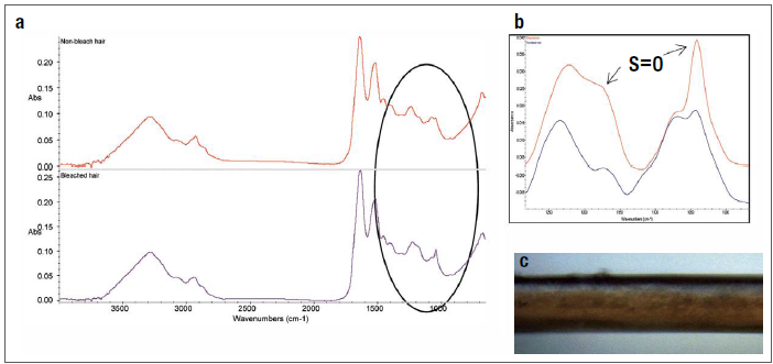
Figure 2. (a) Spectra of non-bleached (red) and bleached (purple) hair. (b) Expanded region showing S=O stretching regions (c) Video image of unbleached hair fiber. Image Credit: Thermo Fisher Scientific - Vibrational Spectroscopy
Visual microscopy is also used to distinguish between natural and synthetic fiber evidence. A highly experienced forensic scientist can recognize the physical properties that distinguish several generic fiber types.
Additional investigation, including chemical analysis, is required to determine the chemical subclass.1 FTIR microscopy has evolved as a powerful analytical method for rapidly determining a fiber's subclass in a non-destructive manner with minimal sample preparation. All this is significant in the forensic community, where preserving evidence is crucial.
Recently, federal money mints have added unique fibers to the paper as an additional barrier against counterfeiting. Figure 3 shows how to inspect the minuscule security fibers in a circulating banknote using attenuated total reflection (ATR) on the Nicolet iN10.
The Nicolet iN10's visual image clearly shows the red fiber, and the ATR data confirms that it is nylon. ATR microspectroscopy yields high spectral quality with minimal cellulose contribution, enabling outstanding library identification.
Figure 3. (Upper spectrum) Spectrum of Nylon fiber embedded in currency. (Lower spectrum) Nylon Spectral library match. (Right) Visual image captured by OMNIC Picta software. Image Credit: Thermo Fisher Scientific - Vibrational Spectroscopy
Tablets
Rapid analytical approaches for determining the chemical composition and distribution of active components for illicit drug tablet analysis are critical in forensic investigations. Sentencing guidelines might be based on both possession and amount, therefore qualitative and quantitative data is required.
Imaging with the Nicolet iN10 MX Infrared Imaging Microscope is a rapid and nondestructive analysis approach suitable for both homogeneous and heterogeneous tablets.
Unlike other macroscopic analytical techniques, FTIR microspectroscopy does not require sample dissolution, which can degrade evidence and result in insoluble or re-crystallized products.
The Nicolet iN10 MX, OMNIC Picta Software, and Thermo Scientific™ OMNIC™ Specta™ Analysis Tools offer drug composition data and insights into the illegal production process. When combined with the system verification tools, the investigator can obtain valuable information for use in court.
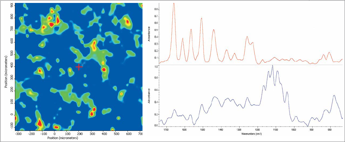
Figure 4. (Left) Chemical image of prescription drug. (Right) Red spectrum is the active ingredient and the blue spectrum is the excipient. Image Credit: Thermo Fisher Scientific - Vibrational Spectroscopy
The Nicolet iN10 MX Imaging Infrared Microscope is the first instrument designed exclusively for chemical imaging analysis, while maintaining the speed, sensitivity, and resolution of conventional infrared microscopy. Figure 4 depicts a chemical image of a prescription drug pill obtained using rapid imaging mapping on a Nicolet iN10 MX.
Infrared data was obtained across a 5 × 5 mm area in approximately five minutes. The chemical image shows the active ingredient in blue; this is the bulk of the material.
However, the green and red contours show that another component is there. Simply clicking within one of the green/red contours shows the spectrum of the second tablet component, which in this case is an unregulated excipient.
The OMNIC Picta Software has automatic collection and analysis wizards. For example, the random mixture wizard may examine and identify many components with a single click. Figure 5 is the multicomponent wizard screenshot for an over-the-counter tablet.
The wizard generates a list of the main components by cross-correlating the collected map spectra. The wizard estimates individual component area contributions and offers semiquantitative distribution data. Each component can then be recognized using spectrum library information, which provides additional chemical information.
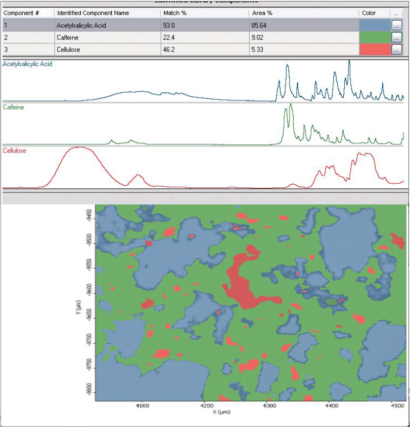
Figure 5. Tablet analysis of an over-the-counter tablet by Picta Multicomponent Wizard. Image Credit: Thermo Fisher Scientific - Vibrational Spectroscopy
Trace analysis
Fingerprint information can help identify or confirm a suspect's involvement in a crime. While fingerprints are unique to an individual, they include more information than just the fingerprint pattern. FTIR microspectroscopic examination can reveal chemical information left behind by fingerprints.
This chemical information can be used to trace a suspect's final steps before committing a crime.
Figure 6 depicts the chemical and video images of a 2 x 2 mm fingerprint impression on a reflecting microscope slide. The primary component of the fingerprint is natural sebum oil from the skin (triglyceride esters).
Some minor curves outside the fingerprint point to another component. The lower right chemical picture in Figure 6 depicts a fingerprint region that contains a small quantity of fibrous wood particles. Chemical imaging instantly determines the unique fingerprint pattern while exposing essential and unexpected trace chemical information.
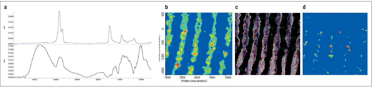
Figure 6. (a) Spectrum of natural triglyceride esters; (b) chemical image of fingerprint; (c) video image of fingerprint; (d) chemical image highlighting the fibrous wood contaminate. Image Credit: Thermo Fisher Scientific - Vibrational Spectroscopy
Paint analysis
Paint chip evidence can be collected at a crime scene involving an automobile. In most circumstances, the paint or paint chip is transferred to a victim or object involved in the collision.
Automotive paint is made up of several layers of chemically diverse materials, such as binders, primers, pigments, and protective resins, which are applied separately to a car's plastic or metal surface.
A chip of paint usually contains information about the different paint layers and can be viewed under an optical microscope. Typically, chemical identification of paint layers necessitates dissolution and chemical extraction.
Fast mapping FTIR microscopy enables immediate chemical identification of each layer. The images in Figure 7 depict the investigation of a multi-layer paint sample. Layer 1 is the outside protective polyurethane coating, layer 2 is the base coat and polypropylene polymer (the bumper's primary component), and layer 3 is the paint binder layer.
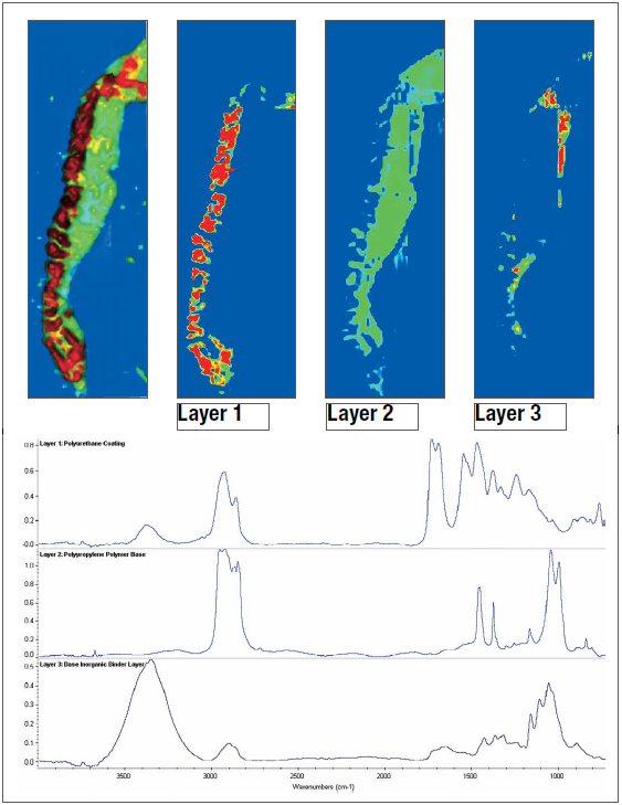
Figure 7. (Upper) Chemical image of a car bumper paint chip layers. (Lower) Spectra of identified layers: Layer 1: protective coating. Layer 2: base coat and polypropylene polymer, Layer 3: binder layer. Image Credit: Thermo Fisher Scientific - Vibrational Spectroscopy
Residues
Chemical residues from crime scenes can provide crucial information and clues. Residues are generally sensitive to evidence handling and should be evaluated with little interaction. FTIR microscopy may detect and evaluate trace contaminants without requiring sample preparation or removal.
Figure 8 shows the sensitivity of infrared microscopy in this application. A fragment of a 10-cent Euro coin was examined with the Nicolet iN10 MX Imaging Microscope. The detailed chemical image (top left) shows a thin pink outline surrounding the stamped coin marks.
The residue spectrum reveals that the material is protein-based and most likely derived from human skin and oil residues. However, this shows how rapidly evidence may be evaluated for trace materials.
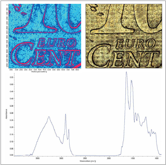
Figure 8. (Upper Left) Chemical image of 10-cent Euro coin (Upper Right) Mosaic video capture of coin sample area (Lower) Spectrum of amide residue. Image Credit: Thermo Fisher Scientific - Vibrational Spectroscopy
Summary
The Nicolet iN10 Infrared Microscope and Nicolet iN10 MX Imaging Microscope provide forensic scientists with immediate visual and chemical information for a wide range of samples. Infrared is sensitive and non-destructive, allowing for correct interpretation while conserving evidence.
The spatial resolution and sensitivity of linear array imaging enable the rapid detection of trace materials. OMNIC Picta's performance verification and validation package provides confidence in the results, which is critical when presenting data in court.
In addition, Thermo Scientific provides unique OMNIC Specta Software, which boasts the most advanced peak and multicomponent search functionality.
References
- Tungol, M.W., Bartick, E.G. and Akbar Montaser (1990). The Development of a Spectral Data Base for the Identification of Fibers by Infrared Microscopy. Applied Spectroscopy, 44(4), pp.543–549. https://doi.org/10.1366/0003702904087451.
About Thermo Fisher Scientific - Vibrational Spectroscopy
With over 50 years of experience in spectroscopy innovation, Thermo Fisher Scientific's vibrational spectroscopy solutions—including near-infrared (NIR), multi-range Fourier transform infrared (FTIR), and Raman instruments—deliver the performance, reliability, and ease-of-use you need to succeed.
Perform raw material identification, differentiate between polymorphs, and analyze formulated products with specificity, speed, and reliability. Analyze biological samples for protein structure elucidation, protein stability, protein-protein interaction, lipid modifications, tissue imaging, analyzing plant extracts, and more with complementary vibrational spectroscopy techniques. With software tools to support data integrity and required validation protocols, and hardware modules designed to work 24-7-365 with minimal re-qualification needs, these spectrometers help you comply with pharmacopeia regulatory requirements while getting the job done.
The Thermo Scientific™ Nicolet™ family of spectrometers are designed to be used in research labs, at-line production, and even at the loading dock to provide the information you need to make critical decisions fast, and with confidence. From drug development and quality control to the identification of the chemical structure of samples and detection of defects and contaminants, Thermo Scientific Raman spectrometers make it easy to characterize molecular structures without becoming a Raman expert.
Sponsored Content Policy: News-Medical.net publishes articles and related content that may be derived from sources where we have existing commercial relationships, provided such content adds value to the core editorial ethos of News-Medical.Net which is to educate and inform site visitors interested in medical research, science, medical devices and treatments.