Sponsored Content by Vitrek LLCReviewed by Maria OsipovaSep 18 2025
GaGe high-performance digitizers are transforming neurological diagnostics and brain research by delivering the speed and accuracy required for sophisticated brain imaging and neurological research. In focused ultrasound therapies, these devices enable instantaneous tracking to open the blood-brain barrier for safe and precise drug delivery.

Image Credit: Vitrek LLC
Moreover, their ultrafast data acquisition supports EEG, MEG, and brain-computer interface applications, delivering precise signal capture for diagnostics and therapeutic development. GaGe technology provides more detailed insights into both brain function and next-generation neurotherapies.
Early detection of ovarian cancer: Three-in-one imaging breakthrough for early detection of a silent killer
Challenge: Detecting ovarian cancer is notoriously difficult in its early stages. Commonly referred to as a “silent killer,” it presents minimal symptoms until reaching an advanced stage—at which point, treatment options are limited and survival rates drop substantially.
Conventional diagnostic instruments lack the resolution, imaging depth, and contrast required to identify malignancies before they spread. Consequently, there is a critical need for a more effective diagnostic solution that enables earlier and more reliable ovarian cancer detection.
How the GaGe digitizer was used: A three-in-one endoscopic probe that integrates optical coherence tomography (OCT), photoacoustic imaging (PAI), and ultrasound (US) provides in-depth, instantaneous imaging for early detection of ovarian cancer. Ultrafast GaGe digitizer cards enable synchronized data capture from all three modalities, guaranteeing comprehensive and precise diagnostic data from a single procedure.

Image Credit: Vitrek LLC
Cracking cataracts: Precision lens hardness mapping with ultrasonic imaging
This paper is a feasibility study of utilizing high-frequency ultrasonic Nakagami imaging for evaluating cataract lenses in vitro.
Challenge: Cataracts cause progressive hardening of the eye’s lens, and successful treatment frequently depends on evaluating the degree of lens hardening. Unfortunately, current preoperative methods lack a dependable, quantitative approach for assessing lens hardness. This forces surgeons to estimate, potentially leading to inefficient energy use during ultrasound treatment and increasing the likelihood of complications.
How GaGe digitizers were used: A 14-bit, 200 MS/s Gage digitizer was used to precisely capture waveforms from a 35 MHz ultrasonic transducer aimed at pig eye lenses. The high-resolution signals allowed Nakagami statistical analysis to precisely analyze lens hardness, demonstrating a possible path to more customized and efficient cataract treatment.
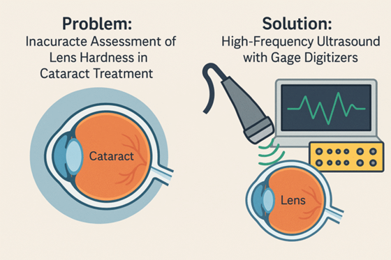
Image Credit: Vitrek LLC
Pulse check: Smart imaging for early artery disease detection
Challenge: Although arterial stiffness is a major indicator of cardiovascular disease, conventional imaging techniques struggle to identify subtle variations in vessel wall features, particularly in early stages. Detecting localized stiffness or inhomogeneity—especially in tiny or complex arteries—requires high-resolution imaging and accurate signal capture, which can be challenging to achieve in both phantom models and live subjects.
How GaGe digitizers were used: Adaptive Pulse Wave Imaging (PWI) was employed to track pressure wave propagation and vessel wall stiffness using 5 MHz and 30 MHz ultrasonic transducers. The transducers were translated across arterial phantoms and mouse aortas to map spatial inhomogeneities in stiffness.
A 14-bit, 200 MS/s Gage digitizer captured high-speed, high-fidelity signals from the ultrasound probes, enabling precise, real-time monitoring of wall motion and stiffness. This method serves as a robust, automated solution for early detection of arterial abnormalities and cardiovascular risk.
Image Credit: Vitrek LLC
Seeing through the needle: High-res imaging for accurate breast cancer detection
Challenge: Breast biopsies are essential in detecting early signs of cancer, including microcalcifications. Traditional ultrasonic imaging used for guiding biopsy needles frequently lacks the accuracy necessary for precise targeting of tiny or ambiguous lesions. This limited imaging resolution can result in false negatives, delaying diagnosis and potentially jeopardizing patient outcomes.
How GaGe digitizers were used: To improve biopsy precision, an innovative method was developed using a miniature 74 MHz ultrasonic transducer embedded directly into the biopsy needle. This high-frequency sensor delivered instantaneous, localized imaging to supplement conventional ultrasound guidance.
A Gage digitizer card captured the rapid, high-resolution transducer waveforms, enabling accurate visualization of tissue structures at the needle tip. This integrated solution substantially enhances targeting precision, lowering the likelihood of missed diagnoses.
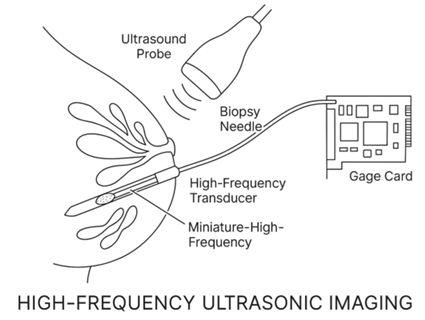
Image Credit: Vitrek LLC
Striking gold: Photoacoustic imaging for stem cell monitoring
Challenge: Stem cell therapies lack dependable in-body monitoring. Although stem cells hold great potential for treating various diseases, a significant limitation remains: monitoring their location and behavior inside the body is challenging once injected. Without a reliable technique for real-time monitoring of stem cells, assessing their efficacy or guiding treatment becomes a substantial obstacle.
How the GaGe digitizer was used: To address this challenge, the research team investigated tagging stem cells with gold nanocages, which are detectable via photoacoustic microscopy. Two in-vitro monitoring techniques were assessed: two-photon microscopy and photoacoustic imaging.
For the photoacoustic technique, a 50 MHz ultrasonic transducer acquired signals from the gold nanocages, while a 12-bit, 200 MS/s Gage digitizer amplified and digitized those signals with high accuracy. This configuration demonstrated a promising path for real-time, non-invasive monitoring of stem cell therapies.
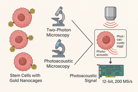
Image Credit: Vitrek LLC
About Vitrek LLC
Since 1990, Vitrek has provided innovative global solutions for high voltage test and measurement including electrical safety compliance testers, multi-point high voltage switching systems and graphical power analyzers. The recent acquisition of MTI Instruments expands their test and measurement portfolio to include non-contact measurement devices, portable signal simulators and calibrators, semiconductor/solar metrology systems and turbine engine/rotating machine balancing. The acquisition of DynamicSignals’ portfolio adds a wide array of board-level data acquisition and integrated real-time RF record/playback system solutions from GaGe, KineticSystems and Signatec. Vitrek also supplies precision high voltage measurement standards to national laboratories and calibration labs around the world. This unique and complementary combination of product and engineering capabilities positions Vitrek as a leading provider of test solutions serving the photovoltaic, medical equipment, power conversion, electrical/electronic component, semiconductor, aerospace and appliance industries. Vitrek is an accredited ISO 17025 Calibration Laboratory.
Sponsored Content Policy: News-Medical.net publishes articles and related content that may be derived from sources where we have existing commercial relationships, provided such content adds value to the core editorial ethos of News-Medical.Net which is to educate and inform site visitors interested in medical research, science, medical devices and treatments.