Working with biological samples that have limited volumes or are potentially inaccessible presents major challenges to achieving actionable biological insights and reliable data in protein biomarker research.
This barrier is present in a diverse array of research domains and includes volume-limited samples, such as ocular matrices, tumor biopsies, or pediatric specimens, as well as samples that necessitate challenging workflows like extracellular vesicles, cerebrospinal fluid, and interstitial fluid.
These sample types typically offer important biological insights into human health and disease, closely reflecting physiological changes. They are also key to sourcing protein biomarkers associated with disease initiation, progression, and treatment response.
The performance of high-throughput protein analysis from minute volumes of these precious sample types has the potential to make a key difference in:
- Understanding pathological mechanisms
- Enhancing patient stratification
- Identifying therapeutic targets
- Expediting drug development
Understanding human disease through studying alternative matrices
A study comparing plasma protein levels with aqueous (AH) and vitreous humor (VH) in patients with varying retinal pathologies offers a clear example of the value of using challenging samples for deeper clinical insights. This study showed that the correlation between the serum and ocular matrices was weak and limited to a few proteins.
A notable portion of the analyzed proteins (∼41 % of detectable proteins) was shown to be correlated between AH and VH, however. This included proteins relevant to retinal pathology, such as immune-related proteins, angiogenesis regulators, and matrix metalloproteinases (Figure 1).
These findings show that AH is linked to considerably fewer vision-threatening complications upon sampling than VH. This could serve as a representative source of biomarker measurements for retinal disease research and monitoring.1
Cytokine profiling of AH also facilitated the identification of three prognostic tumor clusters in uveal melanoma. These clusters differed in terms of patient age and disease stage, highlighting the potential role of these cytokines in the lack of effective antitumor immune responses.2
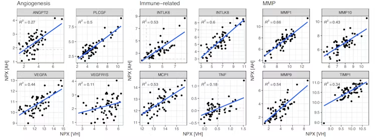
Figure 1. Scatterplots of the protein quantities (NPX) in AH and VH relevant to ocular biology. Blue lines show linear fit. Spearman correlation R2 values are indicated. Image Credit: Olink®- Part of Thermo Fisher Scientific
Although cerebrospinal fluid (CSF) must be obtained through a challenging, invasive collection method, it offers essential insights into the development and progression of neurological diseases.
In another study, proteomic profiling was performed on CSF samples sourced from patients with Parkinson’s disease (PD), progressive supranuclear palsy, corticobasal syndrome, multiple system atrophy, and controls to identify novel diagnostic biomarkers for PD via discovery and validation cohorts.
A total of six proteins were found to be significantly different between PD and controls in both cohorts (including midkine, SMAD5, CCL17, DDC, TFPI-2, and TF). These proteins could serve as potential novel diagnostic biomarkers for PD (Figure 2).3
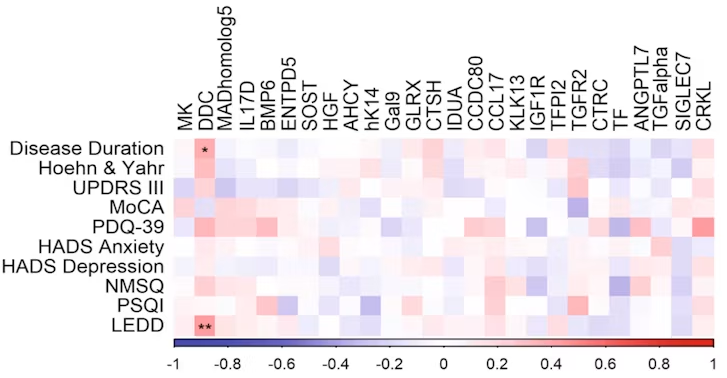
Figure 2. Association of CSF protein expression with PD clinical parameters. Spearman’s rank, adjusted for age and sex, between CSF levels of significantly differentially expressed proteins in PD Stockholm cohort patients and disease duration, scores of Hoehn&Yahr, Unifed Parkinson’s Disease Rating Scale (UPDRS) part 3, Montreal Cognitive Assessment (MoCA), Hospital Anxiety and Depression Scale (HADS), Non-Motor Symptoms Questionnaire (NMSQ) and Pittsburgh Sleep Quality Index (PSQI), and Levodopa equivalent daily dose (LEDD) (BH adjusted P-values; *P<0.05; **P<0.01). Image Credit: Olink®- Part of Thermo Fisher Scientific
Gaining more from limited blood samples
Extracting protein insights from limited sample volumes is particularly essential in studies involving neonatal and pediatric blood.
Human specimen use involves strict regulations when working with pediatric cohorts, often limiting acceptable collection methods, sample volumes, and analyses. However, these samples have proven to be key to identifying novel protein biomarkers for disease diagnosis and prediction in pediatric research.
For example, Dong et al. examined the inflammatory proteins involved in necrotizing enterocolitis (NEC) in a recent study. This severe gastrointestinal disease of the newborn causes high mortality in premature infants.4
Proteomics analysis of plasma samples from newborn infants with sepsis or NEC, as well as healthy controls, revealed 11 inflammatory proteins that could distinguish NEC from controls. These proteins were IL-8, IL-24, TSLP, LIF, OPG, TRAIL, TNFSF14, MCP-4, CCL20, CXCL1, and MMP-10 (Figure 3).
These results offer a new strategy for the early detection of NEC, as combinations of these markers showed significantly improved diagnostic value compared with any individual protein or CRP, a commonly used infection marker.
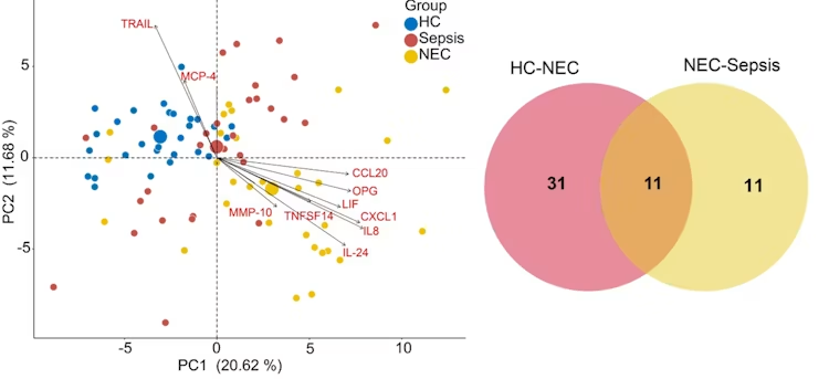
Figure 3. Principal component analysis based on protein levels observed in NEC, sepsis, and HC groups. Wayne diagram highlighting the common and unique proteins in NEC and sepsis groups. Image Credit: Olink®- Part of Thermo Fisher Scientific
Limited blood samples present an issue beyond the sphere of pediatric research. For example, intracranial serum can only be collected in minute volumes, yet it has proven an essential source of insights into ischemic stroke prognostics and diagnostics.
Maglinger et al. demonstrated systemic and intracranial VCAM1 links to stroke comorbidities, functional outcomes, stroke severity, and the role of VCAM1 in molecular signaling pathways.5
These findings could support the prediction of stroke severity, enhance stroke diagnostics and prognostics, and offer practical therapeutic targets for drug repurposing and development.
The proximity extension assay as a solution to limited sample volumes
Challenging sample usage, such as that outlined above, was enabled by Olink’s proximity extension assay (PEA), a robust platform for multiplex protein analysis using just 1 μL of sample.
Its built-in quality control system can be used to monitor each step of the PEA protocol, while only a single measurement is required to ensure high-quality, reliable data.
PEA ensures accurate protein measurements while saving precious sample volume by circumventing the need for replicates. It has also been widely referenced in scientific literature, highlighting its compatibility with a diverse array of sample matrices across key therapeutic areas.
PEA is helping to reveal previously inaccessible protein insights by enabling sampling of substances such as:
- Mouse serum, plasma, and tissue lysate
- Tears, aqueous and vitreous fluid
- Extracellular vesicles
- Cerebrospinal fluid
- Interstitial fluid
References and further reading
- Wilson, S., et al. (2023). Correlation of Aqueous, Vitreous, and Serum Protein Levels in Patients With Retinal Diseases. Translational Vision Science & Technology, 12(11), 9. DOI: 10.1167/tvst.12.11.9. https://tvst.arvojournals.org/article.aspx?articleid=2792990.
- Wierenga, A. P. A., et al. (2019). Aqueous Humor Biomarkers Identify Three Prognostic Groups in Uveal Melanoma. Investigative Opthalmology & Visual Science, 60(14), 4740. DOI: 10.1167/iovs.19-28309. https://iovs.arvojournals.org/article.aspx?articleid=2755740.
- Paslawski, W., et al. (2023). Large-scale proximity extension assay reveals CSF midkine and DOPA decarboxylase as supportive diagnostic biomarkers for Parkinson’s disease. Translational Neurodegeneration, 12(1). DOI: 10.1186/s40035-023-00374-w. https://link.springer.com/article/10.1186/s40035-023-00374-w.
- Dong, H., et al. (2023). Screening inflammatory protein biomarkers on premature infants with necrotizing enterocolitis. Inflammation Research, 72(4), 757–768. DOI: 10.1007/s00011-023-01702-6. https://link.springer.com/article/10.1007/s00011-023-01702-6.
- Maglinger, B., et al. (2021). Intracranial VCAM1 at time of mechanical thrombectomy predicts ischemic stroke severity. Journal of Neuroinflammation, 18(1). DOI: 10.1186/s12974-021-02157-4. https://link.springer.com/article/10.1186/s12974-021-02157-4.
About Olink®- Part of Thermo Fisher Scientific
Olink’s mission is to accelerate proteomics together with the scientific community, to understand real-time biology and gain actionable insights into human health and disease. Our innovative solutions deliver highly sensitive and accurate protein quantification, giving scientists the power to investigate complex biological processes with precision.
One platform. Endless possibilities.
Explore up to 5,400 proteins with high specificity, transparent data, and the flexibility to answer any research question. Meet the next-generation proteomics platform trusted by the scientific community, from small academic research teams through to leading pharma companies.
Sponsored Content Policy: News-Medical.net publishes articles and related content that may be derived from sources where we have existing commercial relationships, provided such content adds value to the core editorial ethos of News-Medical.net, which is to educate and inform site visitors interested in medical research, science, medical devices and treatments.