Sponsored Content by Tescan GroupReviewed by Ify IsiborJun 16 2026
Accessing Intracellular Protein Crystals for Electron Diffraction Using Fluorescence-Guided Cryo-FIB Lamella Preparation
This article describes a workflow for resolving protein structure from crystals grown inside producing cells, based on the recently introduced IncelluloED pipeline, which extends the InCellCryst method of intracellular protein crystal production.1,2
This approach bypasses time-consuming protein purification steps, thereby reducing the number of crystals (ideally to one) required compared with X-ray diffraction methods.
The range of applicable target proteins is also broadened to include those that crystallize with low efficiency, which are otherwise ineligible for untargeted scanning approaches.
The key challenge within this work lies in reliably localizing and targeting potentially rare crystals of interest while ensuring their preparation as electron‑transparent samples from vitrified cells without disrupting the intracellular crystal in situ.
Site-specific cryo-FIB lamella preparation is therefore a key step in ensuring that intracellular crystals are accessible for electron diffraction analysis.
The workflow presented here combines cryo-fluorescence-based localization with targeted cryo-FIB milling using the Tescan AMBER cryo-FIB-SEM system, enabling precise targeting and preparation of lamellae from selected regions of interest.
Challenge statement
Intracellular protein crystals are generally embedded deep within vitrified cellular volumes that are several micrometers thick, placing them beyond the practical penetration depth of electrons in TEM.
Identifying these often rare crystals within crowded cellular environments remains challenging, particularly when precise three‑dimensional positioning is required to avoid missing the target during milling.
At the same time, electron diffraction requires site-specific thinning to lamella thicknesses below ∼300 nm, placing high demands on targeting accuracy and preparation precision.
To address this, fluorescence‑guided approaches enable reliable localization and correlation with cryo-FIB, ensuring accurate transfer of positional information and improving targeting reliability and overall success rates in this application.
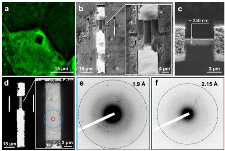
Figure 1: Successfully targeted crystal (a) the original negative fluorescence image of the selected crystal while (b,c) shows SEM and FIB images of the finished lamella containing the same crystal. The crystal is clearly visible on the TEM image in (d) along with diffraction pattern (e) and (f) resulting from fields of view outlined with blue and red circles (Bílá, Pinkas et al. 2026).
Conclusion
Building on these requirements, the fluorescence‑guided, site‑specific cryo-FIB approach presented here provides a practical solution by enabling accurate localization, depth determination, and controlled lamella preparation from selected regions of interest.
By improving targeting accuracy and reproducibility, this workflow significantly increases the success rate of accessing intracellular crystals suitable for electron diffraction analysis.
The full workflow and representative results are described in detail in the complete publication.
References and further reading
- Bílá, Š., et al. (2026). Single-cell structural biology with intracellular electron crystallography. Nature Communications, 17(1). DOI: 10.1038/s41467-026-69205-6. https://www.nature.com/articles/s41467-026-69205-6.
- Schönherr, R., et al. (2024). A streamlined approach to structure elucidation using in cellulo crystallized recombinant proteins, InCellCryst. Nature Communications, 15(1). DOI: 10.1038/s41467-024-45985-7. https://www.nature.com/articles/s41467-024-45985-7.
- Schindelin, J., et al. (2012). Fiji: an open-source Platform for biological-image Analysis. Nature Methods, 9(7), pp.676–82. DOI: 10.1038/nmeth.2019. https://www.nature.com/articles/nmeth.2019.
Acknowledgments
Produced using materials originally authored by Štěpánka Bílá, Dominik Pinkas, Krishna Khakurel, Juliane Boger, Tomáš Bílý, Janos Hajdu, Zdeněk Franta, Roman Tůma, Lars Redecke, and Vitaly Polovinkin.
About Tescan Group
Tescan builds advanced imaging systems that help scientists and engineers explore the micro and nano worlds. In doing so, we help turn observation into insight and questions into progress.
Established by a small team of five engineers in 1991, Tescan has grown into a global company with over 800 employees in 11 countries, united by a brand platform to Accelerate the Art of Discovery. Now Tescan technologies play a central role in laboratories around the world, supporting materials research, failure analysis, and nanoscale imaging with nearly 4500 systems installed in over 80 countries.
Since 2013, Tescan Group has expanded its expertise through a series of acquisitions that have sharpened its technological edge. The merger with ORSAY PHYSICS brought advanced focused ion and electron beam technologies into the Group. In 2018, the acquisition of XRE expanded Tescan’s capabilities in dynamic and micro-CT imaging, opening new possibilities in non-destructive analysis. Another milestone came in 2023. TESCAN ORSAY HOLDING and its subsidiaries were acquired by Carlyle, a U.S.-based private equity firm, marking a new phase of focused investment and global ambition.
In 2024, Tescan Group expanded its technological depth and global footprint. The acquisition of EXpressLO LLC, along with its patent portfolio, added new capabilities in precision stage control and sample handling. That same year, the Group expanded its presence in Asia with the acquisition of TESCAN KOREA Co., Ltd. and DML Co., Ltd. New subsidiaries in Taiwan and Singapore followed, strengthening our commitment to serving scientists where discovery happens.
In 2025, Tescan Group’s commitment to advancing discovery-driven solutions was recognized with an R&D 100 Award in the Analytical/Test category for the TESCAN AMBER X 2, powered by the Mistral™ plasma FIB column.
Tescan Group is headquartered in Brno, Czech Republic, where most systems are designed, assembled, and tested. It’s here that engineering meets purpose and where systems built for discovery are prepared for work in the world’s leading laboratories.
Sponsored Content Policy: News-Medical.net publishes articles and related content that may be derived from sources where we have existing commercial relationships, provided such content adds value to the core editorial ethos of News-Medical.net, which is to educate and inform site visitors interested in medical research, science, medical devices and treatments.