Sponsored Content by TescanReviewed by Ify IsiborJul 15 2026
Biodiversity conservation, botanical research, and ecological studies are all underpinned by accurate taxonomic identification.
Traditional taxonomy tends to rely on visible traits such as flower structure or leaf shape, but these can vary with environmental conditions. Micro-morphological features, however, provide more stable, species-specific markers for reliable classification, particularly features on leaf surfaces.
Among these, leaf trichomes (hair-like appendages) play a dual role, with their morphology and distribution reflecting functional adaptations beyond their taxonomic value.
Surface structures contribute to UV protection, transpiration control, defense against herbivores, and water retention, offering insight into both plant ecological strategies and species differentiation.
However, many diagnostic traits depend not only on individual structures, but also on how micro‑morphological features are distributed across complex plant surfaces. Capturing both their fine detail and their spatial distribution across large areas remains challenging, as conventional optical microscopy lacks the necessary combination of resolution and depth of focus for clear overview imaging.
Tescan MIRA™ SEM overcomes these limitations by providing high resolution and exceptional depth of focus across a wide field of view, enabling researchers to clearly visualize extended plant surfaces while preserving the fine structural details essential for reliable species identification.
Challenge statement
Building on this, accurate species identification increasingly requires not only resolving individual micro‑morphological features, but analyzing their relationships across extended plant surfaces. This places high demands on imaging techniques, which must provide both sufficient detail and consistent image quality over larger areas.
Conventional optical microscopy is limited in this respect, as it cannot deliver the required combination of resolution, depth of focus, and surface sensitivity needed to reliably capture complex epidermal structures in context.
Tescan MIRA™ SEM addresses these challenges by delivering high resolution and exceptional depth of focus across a wide field of view, allowing comprehensive visualization of leaf surfaces while maintaining the detail necessary for taxonomic interpretation.
Low‑voltage SEM imaging further enhances this capability, improving surface sensitivity and enabling accurate representation of delicate plant structures. Combined with appropriate sample preparation, this approach ensures high‑resolution, artifact‑reduced imaging suitable for reliable comparative and taxonomic analysis.
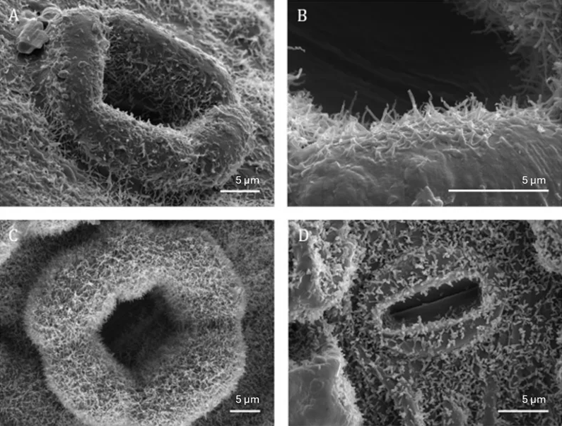
Fig 1. Stomata covered with epicuticular waxes on the abaxial side of the leaves. A, B: Thuja occidentalis, C: Taxus baccata, D: Cotoneaster microphyllus. Image Credit: Mgr. Leona Zuzákova, Masaryk university, Brno.
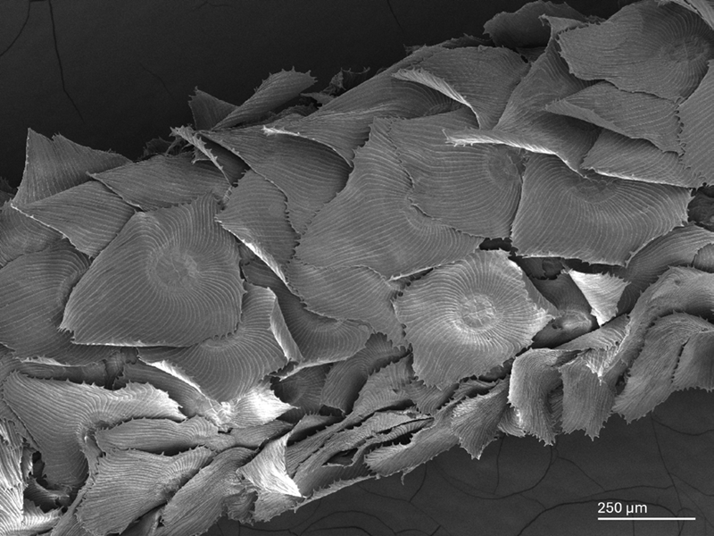
Fig 2. Absorptive trichomes on the leaf surface of Tillandsia ionantha. Image Credit: Mgr. Leona Zuzákova, Masaryk university, Brno.
Methodology
Healthy, mature leaves from specific plant species were collected before being prepared for imaging. Samples were chemically fixed, dehydrated through a graded ethanol series, and then dried using critical point drying (CPD) to minimize structural deformation.
Samples were sputter-coated with a thin gold (Au) or platinum (Pt) layer prior to observation to reduce charging and improve conductivity.
SEM imaging was done using a Tescan MIRA™ microscope across a range of magnifications. This approach allowed the documentation of both overall epidermal organization and fine micro-morphological traits, including stomatal complexes, trichome architecture, and cuticular ornamentation.
Imaging was performed at low accelerating voltages (∼5 kV) to improve surface sensitivity while simultaneously minimizing beam interaction with sensitive biological structures.
The collected images were comparatively analyzed to assess stomatal distribution, trichome density, and cuticular features. This analysis highlighted a series of taxonomically significant differences between the investigated species.
Technology insights
The Tescan MIRA™ offers considerable benefits for high-resolution analysis of plant surfaces. Detailed visualization of microscopic features critical to plant taxonomy is possible, including stomata, trichomes, and cuticular layers.
Key benefits of this instrument include:
- High-resolution imaging: The Tescan MIRA™ offers excellent magnification and clarity, capturing fine plant surface details that cannot be resolved with optical microscopy.
- Wide field navigation: Users can start with a large-area overview at low magnification using Wide Field Optics™, intuitively navigate the sample, and seamlessly zoom in to high-detail imaging without losing structural context.
- Automated imaging routines: Integrated automatic contrast and brightness optimization ensure fast, reproducible image setup with minimal need for manual intervention.
- Fastest time to data: In-Flight Beam Tracing™ minimizes user intervention requirements and ensures consistent acquisition even when operated by inexperienced users.
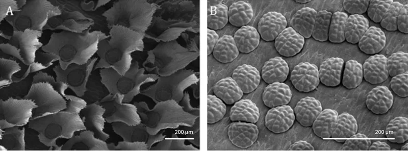
Fig 3. Absorptive trichomes on the leaf surface. A: Tillandsia ionantha, B: Dionaea muscipula. Image Credit: Mgr. Leona Zuzákova, Masaryk university, Brno.
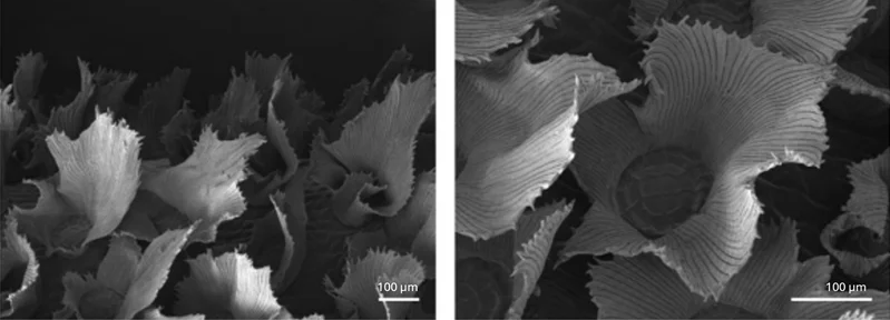
Fig 4. Absorptive trichomes Tillandsia karwinskiana. Image Credit: Mgr. Leona Zuzákova, Masaryk university, Brno.
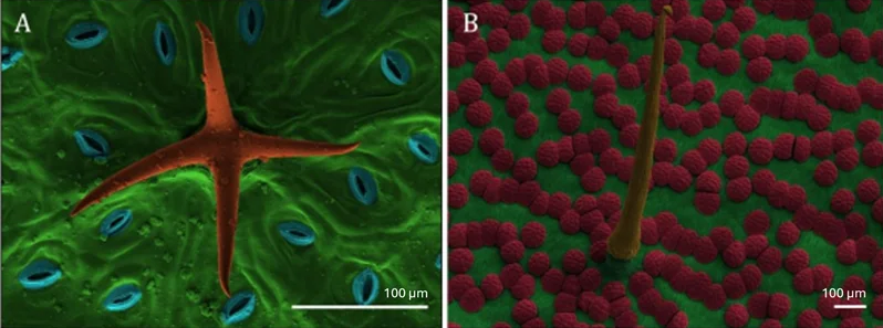
Fig 5. Colorized photographs of plant surface structures: A: Covering trichome on the leaf of Chinese hibiscus (Hibiscus ×rosa-sinensis), B: “Sensitive hair” and digestive glands of the Venus flytrap (Dionaea muscipula). Image Credit: Mgr. Leona Zuzákova, Masaryk university, Brno.
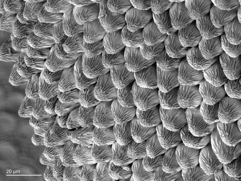
Fig 6. The papillae on the top of flower petals (Rosa centifolia) provide adhesion between the pollinator’s legs and the leaf, which is what makes the pollinator’s landing on the flowers possible. They differ between species. Image Credit: Mgr. Leona Zuzákova, Masaryk university, Brno.
Conclusion
Tescan MIRA™ enables high‑resolution characterization of plant surface micro‑morphology across extended specimen areas, combining clear overview with detailed visualization of structures such as stomata, trichomes, and cuticular ornamentation.
By preserving both spatial context and fine structural detail within a single imaging workflow, it enables confident interpretation of species‑specific traits across large, complex plant surfaces.
This capability supports more accurate taxonomic identification while also providing deeper insight into the functional roles of surface features, including their contributions to protection, regulation, and plant–environment interactions.
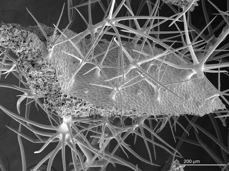
Fig 7. Multicellular trichomes on the leaf surface of Verbascum densiflorum. Image Credit: Mgr. Leona Zuzákova, Masaryk university, Brno.
Acknowledgments
Produced from materials based on the master’s thesis of Mgr. Leona Zuzáková, Photographic Gallery of Selected Plant Surface Structures, defended at the Faculty of Science, Masaryk University in Brno.
About Tescan
Tescan builds advanced imaging systems that help scientists and engineers explore the micro and nano worlds. In doing so, we help turn observation into insight and questions into progress.
Established by a small team of five engineers in 1991, Tescan has grown into a global company with over 800 employees in 11 countries, united by a brand platform to Accelerate the Art of Discovery. Now Tescan technologies play a central role in laboratories around the world, supporting materials research, failure analysis, and nanoscale imaging with nearly 4500 systems installed in over 80 countries.
Since 2013, Tescan Group has expanded its expertise through a series of acquisitions that have sharpened its technological edge. The merger with ORSAY PHYSICS brought advanced focused ion and electron beam technologies into the Group. In 2018, the acquisition of XRE expanded Tescan’s capabilities in dynamic and micro-CT imaging, opening new possibilities in non-destructive analysis. Another milestone came in 2023. TESCAN ORSAY HOLDING and its subsidiaries were acquired by Carlyle, a U.S.-based private equity firm, marking a new phase of focused investment and global ambition.
In 2024, Tescan Group expanded its technological depth and global footprint. The acquisition of EXpressLO LLC, along with its patent portfolio, added new capabilities in precision stage control and sample handling. That same year, the Group expanded its presence in Asia with the acquisition of TESCAN KOREA Co., Ltd. and DML Co., Ltd. New subsidiaries in Taiwan and Singapore followed, strengthening our commitment to serving scientists where discovery happens.
In 2025, Tescan Group’s commitment to advancing discovery-driven solutions was recognized with an R&D 100 Award in the Analytical/Test category for the Tescan AMBER X™ 2, powered by the Mistral™ plasma FIB column.
Tescan Group is headquartered in Brno, Czech Republic, where most systems are designed, assembled, and tested. It’s here that engineering meets purpose and where systems built for discovery are prepared for work in the world’s leading laboratories.
Sponsored Content Policy: News-Medical.net publishes articles and related content that may be derived from sources where we have existing commercial relationships, provided such content adds value to the core editorial ethos of News-Medical.net, which is to educate and inform site visitors interested in medical research, science, medical devices and treatments.