Limitations of Traditional Pathology
Major Multiplex Imaging Technologies
Emerging AI-Integrated Imaging Systems
Applications In Research and Translational Medicine
Bridging The Gap to Clinical Diagnostics
References
Further Reading
Multiplex fluorescence imaging enables simultaneous spatial analysis of multiple proteins and RNA targets within intact tissues, providing a comprehensive view of cellular interactions and tissue architecture beyond conventional pathology. It is increasingly integrating with artificial intelligence and spatial biology workflows to improve biomarker discovery, patient stratification, and clinical decision-making in precision medicine.
 Image credit: Connect Images - Curated/Shutterstock.com
Tissue-based diagnosis has historically depended on the pathologist's ability to interpret stained sections under the microscope. This process, despite its enormous clinical value, captures only a fraction of the molecular information present in a sample. As medicine moves toward increasingly individualized treatment strategies, the demand for deeper, spatially resolved molecular data is fast replacing conventional histological diagnostics.
Multiplex fluorescence imaging (MFI) is providing answers at a depth and spatial scale that conventional diagnostics cannot meet. By enabling simultaneous detection of multiple protein or ribonucleic acid (RNA) targets within a single tissue section, MFI reveals how different cell populations are distributed and how they interact spatially. MFI has also emerged as a foundational technology within the broader field of spatial biology, where molecular measurements are interpreted in their native tissue context to characterize cellular ecosystems and tissue architecture at single-cell resolution.1,2
Each molecular target is labeled with an antibody conjugated to a distinct fluorophore, and fluorescence emissions are separated using optical filters or computational spectral unmixing. Because each fluorophore occupies a different part of the visible spectrum, multiple targets can be imaged within the same tissue section without loss of spatial context.3
Multiplex imaging is rapidly becoming central to cancer diagnostics, enabling the simultaneous characterization of tumor, stromal, and immune cell populations within a single tissue section to inform treatment stratification and patient selection for targeted therapies. In precision medicine, MFI is emerging as an indispensable tool to identify predictive biomarkers and capture the full molecular complexity of the tumor microenvironment (TME).2,4
Download the free PDF to explore how multiplex fluorescence imaging, spatial biology, and AI are transforming the future of precision diagnostics.
Limitations of Traditional Pathology
Standard hematoxylin and eosin (H&E) staining remains important in histopathological diagnosis due to its affordability and reproducibility, but it provides no direct molecular information and relies instead on morphological inference by a trained observer. Although experienced pathologists can infer broad cellular classes from morphology, H&E alone cannot directly identify many clinically relevant molecular phenotypes, immune cell subsets, or protein-expression states.5,10
Single-marker immunohistochemistry (IHC) provides molecular information but also has significant constraints, including the narrow linear dynamic range of chromogenic detection. Moreover, in IHC, when two chromogens colocalize within the same cellular compartment, the resulting mixed signal is difficult to interpret. Similarly, single-plex and low-plex IHC imaging cannot distinguish cell phenotypes defined by the co-expression of multiple markers.2,5
In current diagnostic practice, pathologists review one marker at a time across sequential tissue sections and integrate findings manually. This method is inefficient and prone to inter-observer variability, especially for diagnosing tumors, which require more than a dozen markers for accurate subclassification.5
Moreover, these limitations are particularly apparent when characterizing heterogeneous tissues such as the TME. Predicting response to immune checkpoint inhibition within the TME requires simultaneous, spatially coherent information about numerous cellular functions and protein expression, all of which cannot be provided by a panel of single-plex IHC stains. Multi-marker spatial data are also increasingly needed in neurodegenerative disease pathology, where complex cell-cell interactions within tissue cannot be inferred from single-marker approaches alone.5,6
Major Multiplex Imaging Technologies
Multiplex Immunofluorescence
Tyramide signal amplification (TSA)-based multiplex immunofluorescence has become one of the most widely adopted platforms in translational research. Sequential cycles of antibody staining are followed by horseradish peroxidase-mediated covalent deposition of tyramide-conjugated fluorophores. After each cycle, the antibodies are stripped while the fluorophore signal remains, enabling the process to be repeated with different antibodies.2,4
Some platforms have implemented this principle to achieve six- to eight-plex imaging on formalin-fixed, paraffin-embedded tissue, and the process is compatible with standard clinical auto-stainers, thereby improving the reproducibility of the diagnostic workflow.2,4
Imaging Mass Cytometry
Imaging mass cytometry (IMC) and multiplexed ion beam imaging (MIBI) go beyond fluorescence, using heavy-metal-tagged antibodies detected by mass spectrometry after laser ablation of the tissue. These approaches enable simultaneous detection of more than 40 markers and eliminate issues with fluorophore spectral overlap or autofluorescence.7
The trade-off, however, is substantially lower throughput. The acquisitions from IMC are typically restricted to small regions of interest and require considerable processing time, which makes the clinical use of the method challenging.2,7
CODEX and Spatial Imaging Platforms
Deoxyribonucleic acid (DNA) barcode-based systems, such as CO-Detection by indEXing (CODEX), conjugate each antibody to a unique DNA barcode, which is then detected by complementary fluorescent probes in iterative cycles. This process supports much higher plex counts and detailed immune architecture profiling. The Ultivue InSituPlex technology combines the advantages of spatial imaging platforms and traditional pathology. It allows the same tissue section to be subsequently stained for H&E, which enables the multiplexed fluorescence data to be precisely co-registered with conventional morphological context.2,5,7
 identifies several cell types and molecular markers within a lung cancer tissue sample, while standard H&E staining (centre) reveals the tissue") Combining multiplex fluorescence imaging with traditional pathology. Multiplex fluorescence imaging (left) identifies several cell types and molecular markers within a lung cancer tissue sample, while standard H&E staining (centre) reveals the tissue's overall structure. When the two images are digitally aligned (right), researchers and pathologists can see both the molecular characteristics and anatomical features of every cell simultaneously. This integrated approach supports more accurate tissue analysis, biomarker discovery, and the development of artificial intelligence tools for disease diagnosis. Image credit: Adapted from Wharton KA Jr, et al (2021).
Combining multiplex fluorescence imaging with traditional pathology. Multiplex fluorescence imaging (left) identifies several cell types and molecular markers within a lung cancer tissue sample, while standard H&E staining (centre) reveals the tissue's overall structure. When the two images are digitally aligned (right), researchers and pathologists can see both the molecular characteristics and anatomical features of every cell simultaneously. This integrated approach supports more accurate tissue analysis, biomarker discovery, and the development of artificial intelligence tools for disease diagnosis. Image credit: Adapted from Wharton KA Jr, et al (2021).
Beyond protein imaging, spatial transcriptomic approaches and multiplex RNA imaging technologies are increasingly integrated with multiplex fluorescence workflows, enabling simultaneous assessment of gene expression programs and protein phenotypes within intact tissue architecture.1
Spectral and Hyperspectral Imaging
Spectral unmixing separates the contributions of individual fluorophores at each pixel, compensating for channel bleed-through and tissue autofluorescence to improve signal accuracy. Hyperspectral imaging (HSI) extends this capability by acquiring a three-dimensional hypercube across hundreds of contiguous spectral bands, resolving subtle spectral differences between fluorophores that conventional multispectral approaches may miss.4,7,8
Together, these advances substantially increase multiplexing capacity. For example, excitation spectral microscopy has demonstrated simultaneous imaging of six spectrally overlapping subcellular targets with approximately 1% crosstalk and temporal resolution as high as 10 ms. Similarly, the integration of HSI with microscopes and laparoscopes is further broadening its translational utility.3,8
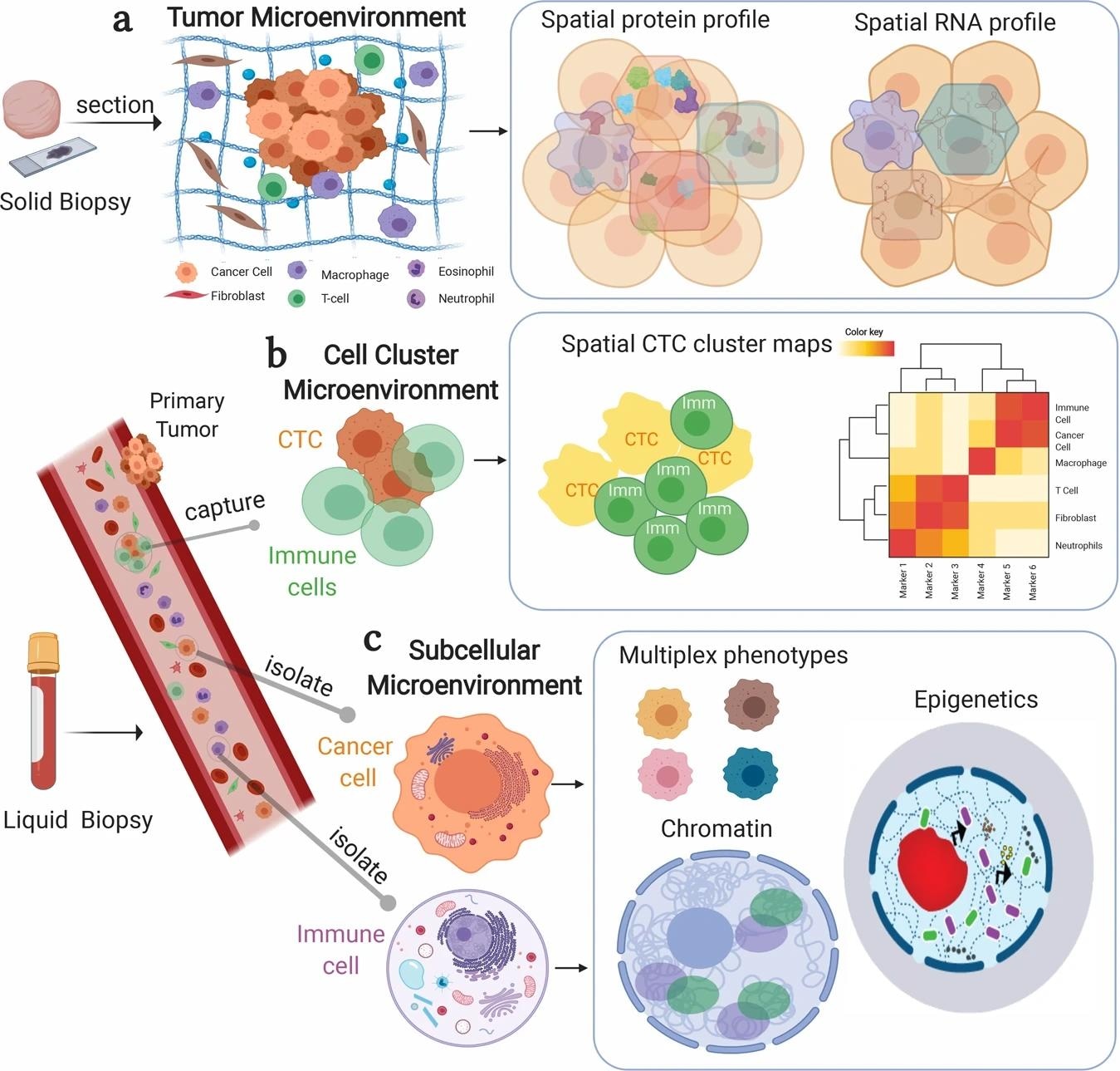 Multiplex imaging reveals disease biology from tissues to individual cells. Researchers can use multiplex imaging to study cancer and other diseases at several levels of detail. In tissue biopsies (top), the technique maps where different proteins and genes are located within the tumor and its surrounding environment. In blood samples (middle), it can identify and analyze clusters of circulating tumor cells and immune cells that may contribute to disease spread. At the single-cell level (bottom), multiplex imaging can examine the internal organization of cells, revealing molecular features that help scientists better understand disease mechanisms, treatment response, and cellular behavior. Image credit: Adapted from Allam M et al, (2020).
Emerging AI-Integrated Imaging Systems
Translating multichannel pixel data into biologically meaningful information requires structured computational pipelines. Machine learning segmentation tools such as Mesmer and Cellpose have substantially improved accuracy over earlier threshold-based methods, particularly in dense tissue samples where cell boundaries are difficult to resolve.7
In label-free imaging, artificial intelligence (AI)-driven virtual staining frameworks employing convolutional neural networks and generative adversarial networks generate multiplexed fluorescence-like outputs from brightfield inputs without exogenous labeling, enabling non-invasive observation of cellular dynamics. Additionally, deep-learning frameworks such as RObust in Silico Immunofluorescence from H&E images (ROSIE) can computationally infer the spatial expression of up to 50 protein biomarkers directly from standard H&E images. ROSIE was trained on more than 1,300 paired H&E and multiplex immunofluorescence specimens spanning multiple tissue types and disease states, demonstrating that computationally generated biomarker maps can identify cellular phenotypes and tissue microenvironments that are not readily discernible from H&E morphology alone.9,10
Automated cell phenotyping via gating or unsupervised clustering, integrated within end-to-end pipelines such as MCMICRO (multiple-choice microscopy pipeline) and PathML, is also enhancing quantitative spatial analysis, with resulting spatial phenotypic signatures demonstrating stronger predictive power for immunotherapy response than single-marker IHC. More broadly, AI models are increasingly being applied to spatially resolved proteomic and transcriptomic datasets for patient stratification, biomarker discovery, and prediction of therapeutic response within precision oncology workflows.1,4,7
Applications In Research and Translational Medicine
In oncology, spatial phenotypic signatures derived from multiplexed imaging are being evaluated as companion diagnostics alongside checkpoint inhibitor therapies. Furthermore, HSI has demonstrated utility across a broad range of cancer types, including cervical, breast, colon, prostate, oral, and head and neck cancers, in both in vivo lesion detection and histological grading. The detailed mapping of immune cell subtypes within the TME is also reshaping our understanding of treatment response.4,5,8
Moreover, the use of multi-marker spatial datasets in neuroscience and infectious disease research is allowing tissue-level characterization of complex cellular ecosystems that single-marker methods cannot resolve. Quantitative image analysis of multiplexed tissue sections is also supporting biomarker validation and mechanistic studies in drug discovery at scale. Emerging spatial proteomic initiatives are generating increasingly large, harmonized molecular datasets that may accelerate biomarker discovery, disease stratification, and target identification across complex diseases.6,11
Bridging The Gap to Clinical Diagnostics
Translating multiplexed imaging into clinical diagnostics requires robust reproducibility across operators, instruments, and sites, as diagnostic decisions, such as patient selection for targeted therapies, depend on consistent test outputs. Preanalytical variables such as ischaemic time, fixation duration, and slide storage conditions substantially affect staining quality and downstream quantification, with an estimated 10–20% of biopsies rendered unfit for analysis due to pre-analytical errors alone.4,7
Data interpretation bottlenecks also persist, as high-dimensional spatial datasets are outpacing manual cognitive capacity, while AI algorithms remain susceptible to artifacts arising from staining variability and tissue heterogeneity. In HSI, the absence of standardized spectral databases limits cross-instrument generalizability. There is also a dearth of standardized biomarker panels and harmonized reference materials, which complicates regulatory submissions and assay calibration. Successful clinical deployment additionally requires cross-site analytical validation, standardized quality-control procedures, harmonized computational workflows, and regulatory frameworks capable of evaluating both laboratory assays and AI-assisted interpretation pipelines.4,5,7,8
Despite these challenges, multiplex platforms are increasingly employed in prospective clinical trials, and as spatial biology develops and evolves, the ability to investigate a biopsy as a complete cellular ecosystem will fundamentally expand the capabilities of diagnostic pathology.4,7
References
- Allam, M., Cai, S., & Coskun, A. F. (2020). Multiplex bioimaging of single-cell spatial profiles for precision cancer diagnostics and therapeutics. NPJ Precision Oncology, 4, 11. DOI:10.1038/s41698-020-0114-1, https://www.nature.com/articles/s41698-020-0114-1
- Harms, P. W., Frankel, T. L., Moutafi, M., Rao, A., Rimm, D. L., Taube, J. M., Thomas, D., Chan, M. P., & Pantanowitz, L. (2023). Multiplex Immunohistochemistry and Immunofluorescence: A Practical Update for Pathologists. Modern Pathology, 36(7), 100197. DOI:10.1016/j.modpat.2023.100197, https://www.sciencedirect.com/science/article/pii/S089339522300197X
- Chen, K., Yan, R., Xiang, L., & Xu, K. (2021). Excitation spectral microscopy for highly multiplexed fluorescence imaging and quantitative biosensing. Light, Science & Applications, 10(1), 97. DOI:10.1038/s41377-021-00536-3, https://www.nature.com/articles/s41377-021-00536-3
- Locke, D., & Hoyt, C. C. (2023). Companion diagnostic requirements for spatial biology using multiplex immunofluorescence and multispectral imaging. Frontiers in Molecular Biosciences, 10, 1051491. DOI:10.3389/fmolb.2023.1051491, https://www.frontiersin.org/articles/10.3389/fmolb.2023.1051491/full
- Wharton, K. A., Jr, Wood, D., Manesse, M., Maclean, K. H., Leiss, F., & Zuraw, A. (2021). Tissue Multiplex Analyte Detection in Anatomic Pathology - Pathways to Clinical Implementation. Frontiers in Molecular Biosciences, 8, 672531. DOI:10.3389/fmolb.2021.672531, https://www.frontiersin.org/articles/10.3389/fmolb.2021.672531/full
- Liampas I. (2025). Special Issue: "New Insights of Biomarkers in Neurodegenerative Diseases". International Journal of Molecular Sciences, 26(22), 10869. DOI:10.3390/ijms262210869, https://www.mdpi.com/1422-0067/26/22/10869
- Omar, M., Fanelli, G. N., Socciarelli, F., Ullanat, V., Puchala, S. R., Wen, J., Chowdhury, A., Valencia, I., Scatena, C., Marchionni, L., Umeton, R., & Loda, M. (2025). Antibody-Based Multiplex Image Analysis: Standard Analytical Workflows and Artificial Intelligence Tools for Pathologists. Laboratory Investigation, 105(10), 104220. DOI:10.1016/j.labinv.2025.104220, https://www.sciencedirect.com/science/article/pii/S002368372500220X
- Lu, G., & Fei, B. (2014). Medical hyperspectral imaging: a review. Journal of Biomedical Optics, 19(1), 10901. DOI:10.1117/1.JBO.19.1.010901, https://www.spiedigitallibrary.org/journals/journal-of-biomedical-optics/volume-19/issue-01/010901/Medical-hyperspectral-imaging--a-review/10.1117/1.JBO.19.1.010901.full
- Li, B., Zhao, Y., Xing, J., Xu, X., Xu, F., & Wen, T. (2026). AI virtual fluorescent staining for label-free, multiplexed live-cell imaging. The Innovation, 101256. DOI:10.1016/j.xinn.2026.101256, https://www.cell.com/the-innovation/fulltext/S2666-6758(26)001256-0
- Wu, E., Bieniosek, M., Wu, Z., Thakkar, N., Charville, G. W., Makky, A., Schürch, C. M., Huyghe, J. R., Peters, U., Li, C. I., Li, L., Giba, H., Behera, V., Raman, A., Trevino, A. E., Mayer, A. T., & Zou, J. (2025). ROSIE: AI generation of multiplex immunofluorescence staining from histopathology images. Nature Communications, 16(1), 7633. DOI:10.1038/s41467-025-62346-0, https://www.nature.com/articles/s41467-025-62346-0
- Imam, F., Saloner, R., Vogel, J. W., Krish, V., Abdel-Azim, G., Ali, M., An, L., Anastasi, F., Bennett, D., Pichet Binette, A., Boxer, A. L., Bringmann, M., Burns, J. M., Cruchaga, C., Dage, J. L., Farinas, A., Ferrucci, L., Finney, C. A., Frasier, M., Hansson, O., … Global Neurodegeneration Proteomics Consortium (GNPC) (2025). The Global Neurodegeneration Proteomics Consortium: biomarker and drug target discovery for common neurodegenerative diseases and aging. Nature Medicine, 31(8), 2556–2566. DOI:10.1038/s41591-025-03834-0, https://www.nature.com/articles/s41591-025-03834-0
Further Reading
Last Updated: Jun 5, 2026