This article and associated images are based on a poster originally authored by Laura Nixon, Andrew McCormack, Olivia Matthews, Sergi Marco, Michaela Macdonald, Ben Kennedy, Darryl Turner, and H. Nikki March, and presented at ELRIG Drug Discovery 2026 in affiliation with Carcinotech Ltd and Concept Life Sciences.
This poster is being hosted on this website in its raw form, without modifications. It has not undergone peer review but has been reviewed to meet AZoNetwork's editorial quality standards. The information contained is for informational purposes only and should not be considered validated by independent peer assessment.

Background
Advanced ovarian cancer is associated with poor long-term survival and frequent development of treatment-resistant disease, highlighting the need for more predictive preclinical models. Conventional 2D culture systems do not adequately recapitulate the structural and biological complexity of the tumor microenvironment, limiting translation of preclinical findings.
Here, we developed reproducible 3D bioprinted ovarian tumor models using ovarian cancer cell lines and fresh patient-derived tumor tissue within biomimetic hydrogels. We evaluated model reproducibility, preservation of tumor microenvironment cell populations, small molecule penetration, and therapeutic response to assess their suitability as physiologically relevant microphysiological systems for preclinical oncology drug discovery.
3D bioprinted tumor workflow

Image Credit: Laura Nixon, et al., in partnership with ELRIG (UK) Ltd.
Patient-derived tumor characterization
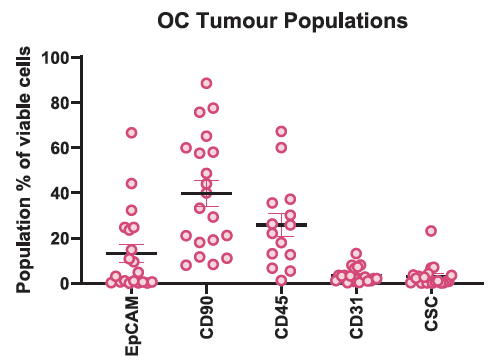
Figure 1. Retention of diverse immune cell populations following dissociation of fresh patient-derived ovarian tumors. Ovarian cancer tissue from treatment-naive and neoadjuvant-treated patients was dissociated and characterized by flow cytometry. Data are shown as each population as a percentage of total viable cells (mean ± SEM), n = 20. Image Credit: Laura Nixon, et al., in partnership with ELRIG (UK) Ltd.
Highly reproducible 3D bioprints
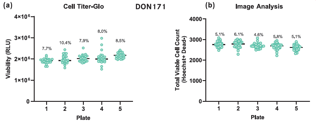
Figure 2. Reproducibility of patient-derived 3D bioprints. Bioprint reproducibility was assessed across five plates generated from a single donor sample (DON 171) using CellTiter-Glo 3D and image-based analysis. Both methods demonstrated low inter-print variability, with coefficients of variation below 10%. Image Credit: Laura Nixon, et al., in partnership with ELRIG (UK) Ltd.
Biological fidelity of 3D bioprinted tumor models
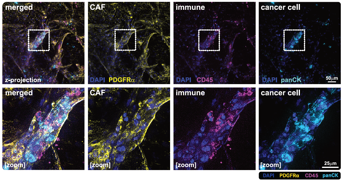
Figure 3. Characterization of patient-derived ovarian cancer bioprints. Patient-derived ovarian cancer tissue was dissociated and bioprinted in a biomimetic hydrogel matrix. After 14 days, bioprints were immunostained for cancer-associated fibroblasts (CAFs, PDGFRα, yellow), immune cells (CD45, magenta), and cancer cells (pan-cytokeratin, cyan), with DAPI nuclear counterstain. Top panels show merged and individual channels; bottom panels show magnified regions. Scale bars: 50 µm (top) and 25 µm (bottom). Images were acquired using a Zeiss Elyra structured illumination microscope. Image Credit: Laura Nixon, et al., in partnership with ELRIG (UK) Ltd.
Small molecule penetration and cellular uptake
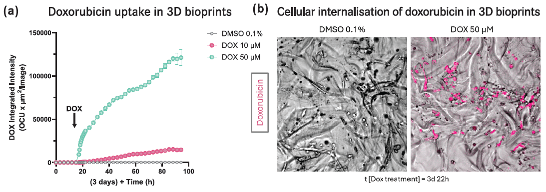
Figure 4. Small-molecule penetration and cellular uptake in patient-derived 3D bioprints. Patient-derived ovarian cancer cells (DON 171) were bioprinted in Px02.28P bioink using the RASTRUM™ platform (Inventia Life Science) and cultured for 7 days. Doxorubicin (10 or 50 µM) was added on Day 4, and uptake was monitored using the Incucyte® SX5 live-cell imaging system. Quantitative image analysis demonstrated dose-dependent doxorubicin accumulation and cellular internalization, supporting efficient diffusion through the bioink and uptake by tumor cells. Image Credit: Laura Nixon, et al., in partnership with ELRIG (UK) Ltd.
Differential drug response in 3D bioprints
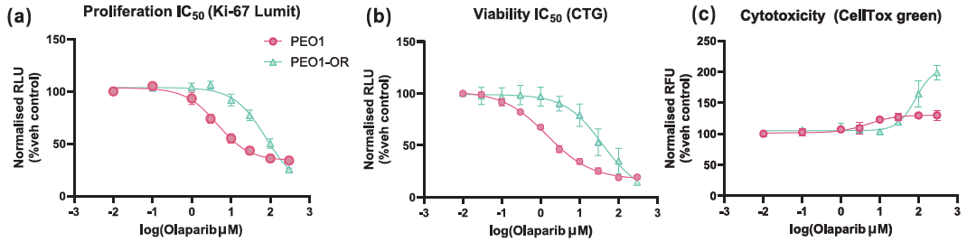
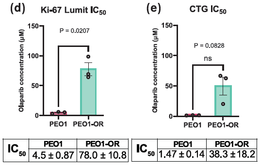
Figure 5. Differential response to olaparib in PEO1 and olaparib-resistant PEO1-OR 3D bioprints. Cells were treated with 0.1–300 µM olaparib for 5 days. (a) Proliferation, (b) viability, and (c) cytotoxicity were assessed using Ki-67 Lumit, CellTiter-Glo (CTG) 3D, and CellTox Green, respectively. IC50 values for (d) CellTiter-Glo 3D and (e) Ki-67 Lumit were compared between models. PEO1-OR bioprints showed reduced olaparib sensitivity. Data are mean ± SEM, n = 3; unpaired t-test with Welch’s correction, P < 0.05. Image Credit: Laura Nixon, et al., in partnership with ELRIG (UK) Ltd.
|
BRCA mutant IC50 |
BRCA restored/WT IC50 |
| Drug |
PEO1 |
UWB1.289 |
PEO1-OR |
OVCAR-3 |
| Carboplatin (µM) |
23.4 ± 5.4 |
23.2 ± 4.0 |
32.8 ± 7.0 |
42.3 ± 2.1 |
| Paclitaxel (nM) |
4.57 ± 1.1 |
n/a |
6.93 ± 2.5 |
1.82 ± 1.6 |
| Olaparib (µM) |
1.47 ± 0.14 |
0.60 ± 0.86 |
38.3 ± 14.8 |
11.0 ± 2.2 |
| Saruparib (µM) |
> 10 µM |
> 1 µM |
> 10 µM |
> 1 µM |
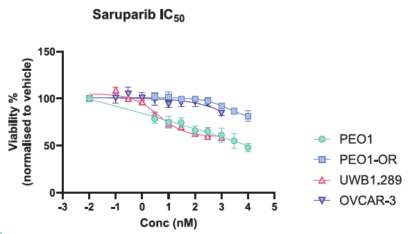
Figure 6. Differential response to saruparib across ovarian cancer 3D bioprints. BRCA–mutant (UWB1.289 and PEO1) and BRCA WT/restored (PEO1–OR and OVCAR–3) models were treated with 0.1–10,000 nM saruparib for 5 days. CellTiter–Glo 3D showed greater reductions in viability in BRCA–mutant models. Image Credit: Laura Nixon, et al., in partnership with ELRIG (UK) Ltd.
Conclusions
- Reproducible 3D bioprinted ovarian tumor models were generated from cancer cell lines and fresh patient-derived tumor tissue, with preservation of key tumor and stromal populations.
- The models enabled quantitative assessment of tumor biology, small-molecule penetration, and therapeutic response within a biomimetic 3D microenvironment.
- Differential responses across molecularly distinct ovarian cancer models demonstrate the ability of the platform to capture biologically relevant variation in treatment response.
- Together, these findings support the use of 3D bioprinted tumor models as physiologically relevant microphysiological systems for translational oncology drug discovery.
About Carcinotech
Carcinotech is a MedTech company with expertise in manufacturing 3D printed living tumors using patient-derived biopsies, primary cells, immune cells and cancer stem cells. Carcinotech’s advanced models provide a platform for rapid, ethical, and accurate drug screening, pre-clinical and personalized medicine testing.
Working with global partners, leading pharma companies, surgeons, pathologists, clinicians, and industry leaders, the goal is to accelerate drug screening and enable the delivery of more effective cancer treatments to market. Access to Carcinotech’s robotic and automated manufacturing allows for accelerated production, with models offering high-throughput capabilities.
About ELRIG (UK) Ltd.
The European Laboratory Research & Innovation Group (ELRIG) is a leading European not-for-profit organization that exists to provide outstanding scientific content to the life science community. The foundation of the organization is based on the use and application of automation, robotics and instrumentation in life science laboratories, but over time, we have evolved to respond to the needs of biopharma by developing scientific programs that focus on cutting-edge research areas that have the potential to revolutionize drug discovery.
Comprised of a global community of over 12,000 life science professionals, participating in our events, whether it be at one of our scientific conferences or one of our networking meetings, will enable any of our community to exchange information, within disciplines and across academic and biopharmaceutical organizations, on an open access basis, as all our events are free-of-charge to attend!
Our values
Our values are to always ensure the highest quality of content and that content will be made readily accessible to all, and that we will always be an inclusive organization, serving a diverse scientific network. In addition, ELRIG will always be a volunteer led organization, run by and for the life sciences community, on a not-for-profit basis.
Our purpose
ELRIG is a company whose purpose is to bring the life science and drug discovery communities together to learn, share, connect, innovate and collaborate, on an open access basis. We achieve this through the provision of world class conferences, networking events, webinars and digital content.
Sponsored Content Policy: News-Medical.net publishes articles and related content that may be derived from sources where we have existing commercial relationships, provided such content adds value to the core editorial ethos of News-Medical.net, which is to educate and inform site visitors interested in medical research, science, medical devices and treatments.
Last Updated: Oct 27, 2026