This article is based on a poster originally authored by Fiona Leslie, Chloe Whiting, and Megan Webster.
TGF-β1-mediated activation of fibroblasts, their transition to α-SMA-expressing myofibroblasts, and the associated increase in the deposition and expression of extracellular matrix proteins represent key pathologic mechanisms of fibrosis.
Modeling this process in vitro enables the evaluation of potential anti-fibrotic therapeutics for their capacity to reduce or reverse fibroblast-to-myofibroblast transition (FMT).
The development and validation of robust assay systems require substantial time and effort.

Image Credit: Newcells Biotech
Newcells Biotech’s team has worked diligently to overcome the many technical challenges to optimize their high-throughput, high-content imaging FMT assay. This article highlights several key difficulties and the corresponding resolutions.
Optimization of plate coatings
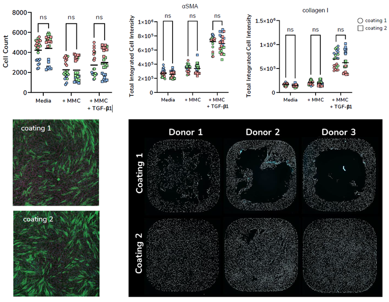
Image Credit: Newcells Biotech
To guarantee consistent cell adherence to culture wells throughout the FMT assay process, including during mechanical washing, cell seeding densities and culture media were optimized to control cell proliferation.
After observing significant cell detachment as a result of mechanical washing, various plate coatings were compared to enhance fibroblast attachment and reduce assay variability. Further work was conducted to optimize immunocy-to-chemical detection of α-SMA and collagen 1.
Optimization of culture media
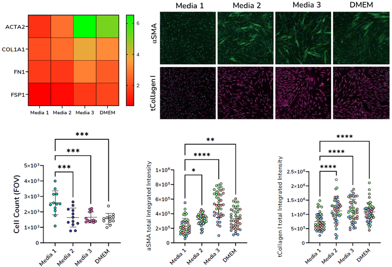
Image Credit: Newcells Biotech
Ensuring a consistent and substantial assay window is vital for establishing a reproducible in vitro assay for compound screening.
Assay culture media was optimized to promote the activation of primary fibroblasts following stimulation with TGF-β1. As shown in Figure 2, Media 3 promoted the expression of both α-SMA and collagen 1 at the gene and protein levels.
Together with refined segmental analysis masks, one can accurately detect changes in the deposition and expression of α-SMA and collagen 1 in response to treatment with potential therapeutic compounds.
Quantification of protein expression
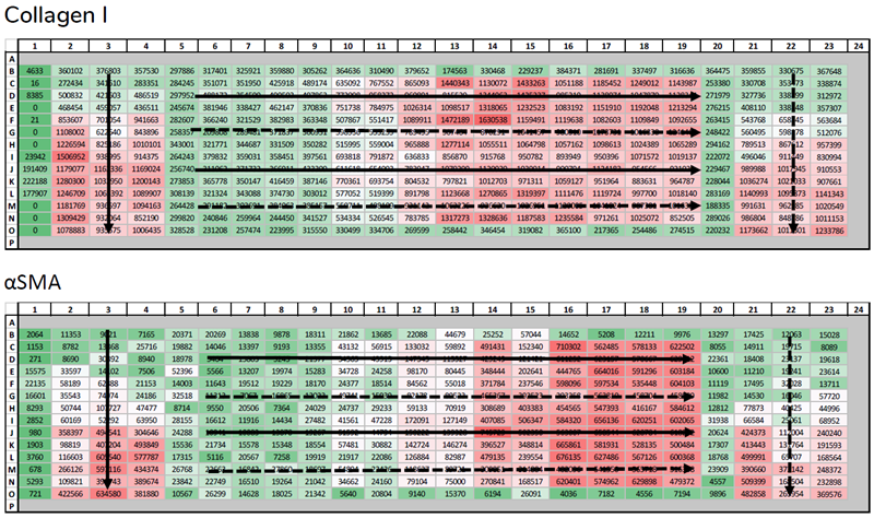
Image Credit: Newcells Biotech
To guarantee consistent data, independent of well position on the plate, α-SMA and collagen 1 were quantified after the stimulation of primary fibroblasts with increasing concentrations of TGF-β1 across both horizontal and vertical axes (arrows).
The effects of two different stimulation methods were also tested (dashes). Applied conditional formatting indicates increased expression of α-SMA and collagen 1, ranging from green to red, respectively.
Newcells Biotech’s FMT assay
Newcells’ FMT assay utilizes high-content imaging to assess the effects of potential therapeutics on fibroblast activation, collagen expression, and deposition in a high-throughput format.
Assay features:
- Three validated primary HLF donors
- High-throughput 384-well format
- Six-point dose response for up to six TAs per plate
- Six technical replicates per condition
- Validated experimental assay controls
Available assay readouts:
- Cell Number
- αSMA
- Collagen I
Acknowledgments
Produced from material originally authored by Fiona Leslie, Chloe Whiting, and Megan Webster from Newcells Biotech.
About Newcells Biotech
Newcells Biotech develops in vitro cell-based assays for drug and chemical discovery and development.
Using our expertise in induced pluripotent stem cells (iPSCs), cellular physiology, and organoid technology, we build models that incorporate the “best biology” for predicting in vivo behavior of new drugs.
Our experts have developed and launched assays to measure transporter function, safety, and efficacy in a range of cell and tissue types, including kidney, retina and lungs.
We have the capability to develop and implement protocols to measure cilia beat frequency and toxicity on small airway epithelial cells model, retinal toxicity and disease modelling on retinal organoids and retina epithelium, as well as drug transport in the kidney, DDI and nephrotoxicity across human and a range of preclinical species.
Sponsored Content Policy: News-Medical.net publishes articles and related content that may be derived from sources where we have existing commercial relationships, provided such content adds value to the core editorial ethos of News-Medical.Net which is to educate and inform site visitors interested in medical research, science, medical devices and treatments.
Last Updated: May 13, 2025
 Newcells Biotech strengthens executive and US team with key appointments