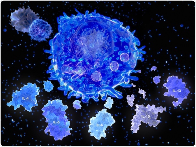
Cytokines play a critical role as mediators of immune responses and are generated by a variety of cells. Methods that can be used to measure cytokine levels in a sample and differentiate between different types are therefore essential tools for understanding the human immune system.
Various bulk bioassays and techniques have been developed to measure cytokine levels. However, all of these assays are based on the assumption that all cells of a given phenotype produce similar levels and types of cytokines. This often leads to ambiguous results and makes it difficult to determine the presence of contaminating cells in the culture. It was for these reasons that there was an extensive search for a method that could identify single cells by using their cell surface markers and cytokine production profiles.
Identifying the cytokines present inside a cell usually presents a problem due to weak intracellular signal. However, a 1993 study tried to circumvent these issues through a novel method that is now widely used for the detection of cytokines IL-1α, IL-6, IL-8, and TNF-α.
 Juan Gaertner | Shutterstock
Monensin to the rescue
Monensin is a carboxylic ionophore that interrupts the intracellular transport process. It binds to Na+, K+, and H+ ions, disrupting the ion gradients in biological membranes. This perturbs the Golgi complex and transport to the cell membrane without interrupting the protein synthesis, subsequently leading to the accumulation of cytokines in the Golgi complex and signal-to-noise ratio increase.
While performing the experiment, human peripheral blood mononuclear cells were activated with phorbol myristate acetate (PMA) and ionomycin. This was done in the absence of monensin in controls and the presence of monensin in test cells. Such treatment increased the signal-to-noise ratio by increasing the intensity of fluorescence.
After 10 hours of stimulation, 50% lymphocytes tested positive for interleukin-2 (IL-2) post monensin use, while only 11% of the cells tested positive of IL-2 in its absence. Also, interleukin-4 (IL-4) producing cells in the peripheral blood of humans are rare, but they could be detected using monensin. However, the mean fluorescence intensity of IL-4 was less compared to the intensity of IL-2, which could be expected if natively their numbers are lower when compared to IL-2.
Determining the optimal concentration of monensin
Adding high doses of monensin could lead to toxicity, thus the optimal dosage for monensin needs to be determined. For this, monensin of varying concentration (from 10 nM to 100 µM) has been used and the percentage of dead cells has been observed to determine the toxicity level.
The results showed a marked level of toxicity at 100 µM that was present even after six hours of culture, and concentrations below 1 µM did not lead to cell death. Also, when used at 1 µM, there was a high degree of toxicity after 24 hours, and after 48 hours almost all cells were dead. The addition of monensin did not alter cell surface markers at both six hours and post-experiment.
Detecting a restricted population using three-color flow cytometry
To assess the levels and type of cytokine population in a subset of cells, earlier it was required to isolate and measure the cytokine levels using ELISA, bioassay or the expression of mRNA. In the aforementioned study, the researchers tested if they could analyze the cytokine pattern in a restricted population of cells using flow cytometry.
Previous studies show that different cells, such as human naïve or memory cells secrete different kinds of cytokines. The cells were initially fixed using a combination of paraformaldehyde (PFA) and saponin, as these do not alter the scatter properties in contrast to Tween 20 and Triton-X. Using three-color FACS the researchers could find differences in the expression of IL-2, IFN-y, and IL-4 in memory and naïve T cells.
Method validation
To validate the results, the researchers performed microscopy along with flow cytometry for ten samples and tested the levels of IL-2, IFN-y, and CD45. They found that the microscopy results were highly correlated with the results from flow cytometry.
The paper describes a relatively quick, easy and sensitive method involving 2-6 hours of culture and 2-3 hours of staining to detect the levels of cytokines. This method required only a few cells (105 cells) in every test.
Also, the cells stained for various cytokines can be stored for 1-3 days in the dark. Thus, this method provides a means to characterize the cytokines in a heterogeneous population, screening cytokine patterns, and functionally characterizing the cytokine-producing cells.
Source
Jung et al (1993) Detection of intracellular cytokines by flow cytometry. Journal of Immunological Methods (https://www.sciencedirect.com/science/article/pii/0022175993901584)
Further Reading
Last Updated: Jul 31, 2019