Tumors consist of a heterogeneous mixture of malignant and normal cells in the parenchyma, fenestrated with an abnormally structured stroma. The inter-patient, and even inter- or intra-tumor, variability expressed by all types of cancer makes the design and testing of medications a challenging prospect.
Predicting the efficacy and toxicity of cancer therapies is key to establishing new drug leads and designing treatment regimens, so researchers have developed a variety of cancer models that allow each aspect to be analyzed at the in vitro and in vivo levels.
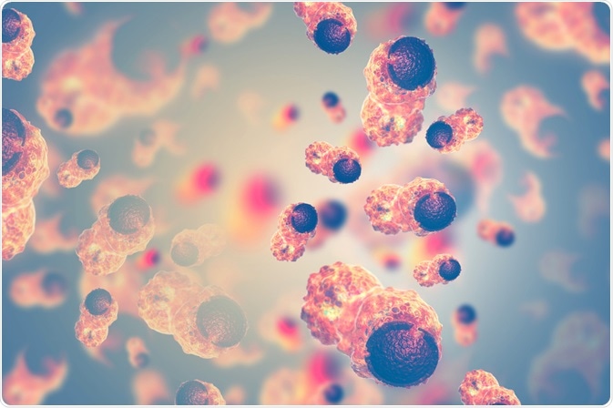
 Cancer cells. Image Credit: crystal light / Shutterstock.com
In vitro cancer models
Oncogenes are genes that have the potential to cause cancer if allowed to run out of control, usually because they control for functions of the cell such as growth or mitosis. Tumor suppressor genes have the opposite function, being involved in tasks such as inducing controlled apoptosis. Mutations on one or both of these types of genes leads to uncontrolled cell growth, cancer.
The unstable nature of the genome of the cancer cell quickly generates progeny with additional mutations through increased proliferation, reduced transcription checking, and a disorganized epigenome. Natural selection is at play amongst the varied population of cancer cells, with those most successful at propagating and adapting to the local microenvironment becoming more populous in that area.
Maintaining this environment while testing the efficacy of drugs against a representatively heterogeneous group of cells has been the primary aim of researchers developing preclinical cancer models.
In vitro cancer cell cultures have commonly been employed in drug testing scenarios for many decades thanks to their reliability, low cost, and ease of setup. These cell cultures are allowed to multiply to confluency before a known number of cells is placed in a 2D well and a known quantity of drug is added.
The remaining percentage of viable cells after a set period of time is direct evidence of the cytotoxicity of a drug, allowing researchers to determine which may be the most efficacious and potent. Combining other analytical techniques with this 2D cell culture method can help to identify the way in which the drug enters the cell and subsequently and interacts to cause the observed effects. However, 2D cell cultures do not capture the true environment observed in vivo, and so cannot be used to reliably infer a significant amount of information regarding how a tissue may react to a drug in a three-dimensional environment.
3D cultures attempt to maintain the cell-to-cell contact found in vivo while also replicating other aspects of the tumor microenvironment such as the stroma, and are sometimes called organoids. These cultures are usually made by taking a biopsy from a patient and growing the tissue in an enriched medium, where they grow in three dimensions and replicate the original tissue architecture with aid from scaffolding.
Modern microfluidics allows the flow of medium around and through the organoid to closely replicate a dynamic in vivo environment. Organoids may take many months to prepare and are difficult to cultivate in large numbers, so an alternative method of maintaining the three-dimensional structure of a tumor is to collect organotypic tumor slices.
Organotypic tumor slices are generated from biopsy samples that are cut into slices of around 200 µm in thickness. The slices are then cultured individually or in small groups, maintaining the native tumor environment for periods of weeks, during which testing can be performed. Cellular heterogeneity is maintained by this method, along with other aspects of the tumor microenvironment and extracellular matrix that organoids fail to replicate due to the bias generated by quickly proliferating cancer cells.
A comparison of the two methods by Sivakumar et al. (2019) noted that both exhibited similar responses to most drugs applied, with one notable exception being the greater sensitivity displayed by tumor slices to vascular endothelial growth factor receptor inhibitor drug vandetanib. This drug acts by limiting angiogenesis, explaining the lessened effect against the organoid model, as it targets the endothelial cells found in the tumor slice only.
In vivo cancer models
While in vitro testing allows for high-throughput screening of drugs, the full complexity of a living system remains difficult to replicate. Modern high-end in vivo testing may take a biopsy from a patient that is then implanted subcutaneously onto a living animal. Using biopsy material maintains the structure and gene expression of the original tumor, allowing researchers to examine the effects of a drug both on the tumor and the living host.
3D Cell Models - Cancer
Biomarkers
The identification and tracking of key biomarkers is a powerful tool in preclinical modeling. Depending on the specific type of cancer, taking into account the mutations that have led to uncontrolled growth and the location of the tumor, a variety of molecules may be produced or overexpressed by the cells. Biomarkers may be in the form of genes, proteins, or small molecules, and each provides information that allows for the diagnosis and prognosis of cancers.
Tracking these biomarkers over the course of in vitro or in vivo experiments using techniques such as fluorescence microscopy supplies information regarding cellular dynamics and the mechanisms at play. This is advantageous over a simple reading of cell viability or tumor mass (in the case of in vitro or in vivo testing, respectively), as factors affecting the mechanism of action can be identified.
Biomarkers that are useful in quantifying various aspects of a cell's function and response to a drug are constantly being sought and discovered, and have become invaluable in predicting efficacy and safety.
In silico cancer models
The identification and tracking of biomarkers, along with other experimental parameters gathered by in vivo and in vitro testing, generates vast quantities of data that can be used to construct in silico preclinical cancer models. The identity of a target site is key information in the development of a drug, and once identified in silico methods can be used to test the affinity of many millions of compounds against the target in a high throughput manner. Computational modeling of the pharmacokinetics and pharmacodynamics of drugs has played a major role in lead discovery in the past several decades, and increasingly detailed and complex simulations are allowing systemic effects to be investigated, and any emergent properties described.
Sources
- Liu, J., Dang, H. & Wang, X. W. (2018) The significance of intertumor and intratumor heterogeneity in liver cancer. Experimental & Molecular Medicine, 50.https://www.nature.com/articles/emm2017165
- Dhandapani, M. & Goldman, A. (2017) Preclinical Cancer Models and Biomarkers for Drug Development: New Technologies and Emerging Tools. Journal of Molecular Biomarkers and diagnosis, 8(5). https://www.ncbi.nlm.nih.gov/pmc/articles/PMC5743226/
- Li, X., Valadez, A. V., Zuo, P. & Nie, Z. (2014) Microfluidic 3D cell culture: potential application for tissue-based bioassays. Bioanalysis, 4(12). https://www.ncbi.nlm.nih.gov/pmc/articles/PMC3909686/
- Sivakumar, R. et al. (2019) Organotypic tumor slice cultures provide a versatile platform for immuno-oncology and drug discovery. Oncoimmunology, 8(12).https://www.ncbi.nlm.nih.gov/pmc/articles/PMC6844320/
Further Reading
Last Updated: Feb 26, 2021