Introduction: Finding Weak-Binding Fragments with NMR
The Challenge of Turning NMR Spectra Into Reliable Hits
How Automated NMR Analysis Changes the Screening Workflow
What Automation Could Mean for Fragment-Based Drug Discovery
Toward More Scalable NMR Screening
References and Further Reading
Automated processing is helping overcome a major bottleneck in NMR-based fragment screening by converting complex spectral datasets into reliable hit lists more efficiently. Advances in 1H and 19F approaches are improving screening throughput while preserving the experimental sensitivity needed to identify weak-binding fragments.
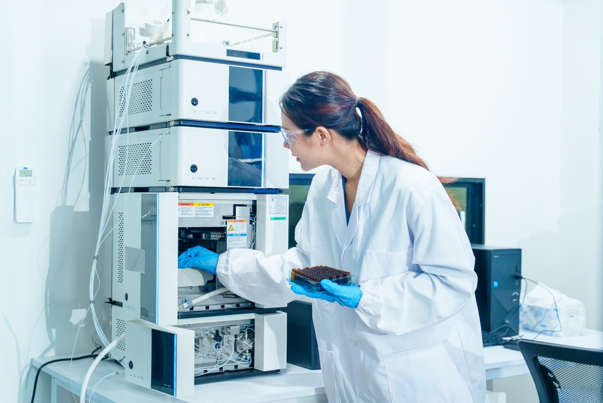 Image credit: Rawpixels stock/Shutterstock.com
Fragment-based drug discovery (FBDD) is a relatively recent pharmacological (drug discovery) advancement that allows researchers to identify low-molecular-weight compounds or ‘fragments’ that bind weakly but specifically to therapeutic targets. While several technologies have been previously used to identify and isolate fragment hits, ligand-observed nuclear magnetic resonance (NMR) spectroscopy is an established and widely used tool for detecting weak fragment–target interactions.1,2
Unfortunately, the large number and complexity of spectra generated during NMR fragment-screening campaigns can make conventional manual analysis a significant FBDD challenge. Researchers addressed these throughput limitations by developing novel, fully automated NMR data-processing modalities that significantly accelerate hit identification without compromising accuracy.1
This article synthesizes the latest peer-reviewed evidence on these advanced automated workflows, while also considering fragment-library quality control, fluorine NMR, and adaptations required for challenging membrane-protein targets.2,3,4
Introduction: Finding Weak-Binding Fragments with NMR
FBDD represents one of the most systematic and targeted approaches to modern drug discovery, specifically lead generation. The process’s underlying principle relies on small, low-affinity compounds to serve as high-quality starting points for downstream pharmacological optimization.1
The small size and transient interactions of these fragments (typical dissociation constants [KD] in the micromolar to millimolar range) necessitate the use of sensitive biophysical techniques for compound detection. NMR spectroscopy is particularly suitable for detecting and characterizing these weak binding interactions and can also be used to determine dissociation constants.1,2
Ligand-observed NMR can detect weak binding events while also providing an internal quality-control check on compound integrity and concentration directly from the screening sample.1
NMR-based FBDD originated with the “SAR by NMR” approach and has subsequently developed into multiple ligand- and protein-observed screening strategies. Ligand-observed methods are particularly useful for primary screening because they do not require isotopically labeled protein and are not intrinsically restricted by receptor molecular size.1,2
While this sensitivity enables novel drug discovery, the manual visual inspection of hundreds to thousands of complex NMR datasets can be tedious, time-consuming, and susceptible to analysis errors, necessitating the development of automated workflows.1
The Challenge of Turning NMR Spectra Into Reliable Hits
Ligand-observed NMR mechanistically identifies interacting compounds by monitoring changes in the NMR signals of fragments upon the introduction of a target protein.
Notably, while different ligand-observed NMR deployments leverage divergent techniques for their respective analytical assays (the most commonly used examples include saturation transfer difference [STD], Carr–Purcell–Meiboom–Gill [CPMG] relaxation-edited experiments, and T1ρ sequences), all these methodologies capitalize on the rapid chemical exchange between the free and bound states of a ligand and the observed transfer of the altered magnetic relaxation properties of the large macromolecular target to the small molecule.1
Furthermore, reviews of proton (1H) and fluorine (19F) detection assays emphasize that these modalities demonstrate unique analytical properties. Consequently, the specific choice of detection assay used is informed by the study’s screening requirements (e.g., sensitivity and mixture composition) and, in turn, dictates the study’s assay design.1
Peng and colleagues (2016) compared the utility and performance of these distinct detection assays and found that proton-detected NMR offers high sensitivity for binding but is typically limited to smaller mixture sizes of 6 to 12 compounds (due to high spectral complexity and significant peak overlap). Furthermore, 1H assays are frequently confounded by solvent interference and signal crowding, both of which have traditionally required expert-driven manual review to control for.1
Conversely, Buchholz & Pomerantz's (2021) review of 19F fluorine-detected NMR advancements found that these methodologies provide very high sensitivity. 19F is a 100%-naturally-abundant nucleus with a gyromagnetic ratio close to that of 1H, giving it about 83% of 1H NMR sensitivity.3
19F NMR implementations can therefore accommodate significantly larger mixture capacities (up to 30 compounds) with low spectral complexity, as there are no biological background signals.1,3
However, 19F NMR assays demonstrate high sensitivity to changes in the local chemical environment, and local peak misalignments can occur between reference and screening spectra.1,3
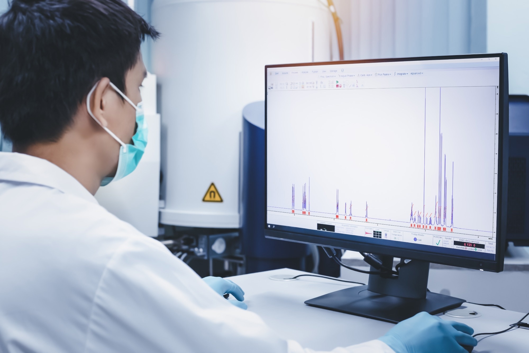 NMR spectra provide detailed information for detecting and characterizing fragment interactions, while automated data-processing approaches can reduce the burden associated with manual spectral interpretation. Image credit: S. Singha/Shutterstock.com
How Automated NMR Analysis Changes the Screening Workflow
Today’s analytical bottlenecks in manual spectrum interpretation are being addressed through the development and validation of fully automated computational procedures to process and analyze both 1H- and 19F-detected ligand-observed NMR binding data.1
Unlike their manually driven traditional counterparts, these automated approaches perform batch processing, spectral alignment, and spectral normalization using predefined templates.1
Peng et al.’s (2016) computational approach, for example, relies on a global spectral deconvolution algorithm to identify and match peaks between reference and screening spectra (e.g., in 1H-detected T1ρ and STD experiments). The software further employs a mathematical tolerance function to compensate for local peak misalignments.1
In 19F-detected CPMG assays, similar algorithms are used to match peaks between blank and protein-inclusive samples using specialized integration regions to account for peak shifts between samples.1
Rather than acting as a computational "black box," Peng et al’s (2016) software links the automated results to an interactive graphical interface, thereby allowing scientists to rapidly visualize candidate binders, review overlapping signals, and verify the associated spectra.1
What Automation Could Mean for Fragment-Based Drug Discovery
The ongoing transition of traditional manual NMR screening workflows to automated analysis is already demonstrating substantial advantages for pharmaceutical and academic drug-discovery laboratories. Automated processing can generate high-confidence hit lists in a fraction of the time required for manual analysis, reducing a significant throughput limitation in ligand-observed NMR campaigns.1,2
This acceleration allows pharmacological laboratories to practically assess larger, more diverse chemical libraries and iterate through follow-up chemistry optimization cycles with shorter analytical turnaround times.1
Automation does not eliminate the experimental constraints of FBDD. Library quality, compound solubility, sample composition, target stability, spectral overlap, nonspecific interactions, and the physicochemical environment of challenging targets can all influence whether a detected spectral response represents useful fragment binding.1,2,4
Toward More Scalable NMR Screening
This article exemplifies how integrating automated NMR analysis into FBDD-based drug-discovery pipelines can significantly mitigate persistent logistical bottlenecks in experimental drug discovery.
Modern NMR-based FBDD is therefore best viewed as an integrated workflow rather than simply an automated hit-detection step: fragment-library quality control establishes that suitable compounds enter the screen; ligand-observed methods provide sensitive and scalable detection of weak binding; protein-observed experiments can provide site-specific information; 19F NMR expands the available screening strategies; and computational analysis increases the throughput with which the resulting spectra can be interpreted.1,2,3,4
By combining high-throughput NMR acquisition with automated data analysis and appropriate experimental validation, researchers can maintain the sensitivity required for FBDD while operating at greater screening throughput.1
References and Further Reading
- Peng, C., et al. (2016). Fast and Efficient Fragment-Based Lead Generation by Fully Automated Processing and Analysis of Ligand-Observed NMR Binding Data. Journal of Medicinal Chemistry, 59(7), 3303–3310. DOI:10.1021/acs.jmedchem.6b00019, https://pubs.acs.org/doi/10.1021/acs.jmedchem.6b00019
- Li, G. C., Castro, M. A., Ukwaththage, T., & Sanders, C. R. (2024). Optimizing NMR fragment-based drug screening for membrane protein targets. Journal of Structural Biology: X, 9, 100100. DOI:10.1016/j.yjsbx.2024.100100, https://www.sciencedirect.com/science/article/pii/S2590152424000059
- Buchholz, C. R., & Pomerantz, W. C. K. (2021). 19F NMR viewed through two different lenses: ligand-observed and protein-observed 19F NMR applications for fragment-based drug discovery. RSC Chemical Biology, 2(5), 1312–1330. DOI:10.1039/d1cb00085c, https://pubs.rsc.org/en/content/articlelanding/2021/cb/d1cb00085c
- Sreeramulu, S., et al. (2020). NMR quality control of fragment libraries for screening. Journal of Biomolecular NMR, 74(10–11), 555–563. DOI:10.1007/s10858-020-00327-9, https://link.springer.com/article/10.1007/s10858-020-00327-9
Last Updated: Oct 8, 2026